Method for detecting uridine monophosphate modification of protein

A technology for the detection of uridine monophosphate and its detection method, which is applied in the field of detection of protein monophosphate uridine acid modification, which can solve the problems that have not received attention and the modification detection method has not been established, and achieve the effect of high sensitivity

Inactive Publication Date: 2020-08-07
INST OF BASIC MEDICINE OF SAMS
View PDF4 Cites 0 Cited by
  • Summary
  • Abstract
  • Description
  • Claims
  • Application Information

AI Technical Summary

Problems solved by technology

Among them, protein uracil mononucleotide modification (UMPylation, UMPylation) was first identified in Escherichia coli in 1975, but the substrate protein of UMPylation was limited to the glutamic acid...

Method used


Image

  • Method for detecting uridine monophosphate modification of protein
  • Method for detecting uridine monophosphate modification of protein
  • Method for detecting uridine monophosphate modification of protein

Examples


Embodiment Construction

[0024] The present invention will be further described according to the following examples, and the mode of the present invention includes but not limited to the following examples.

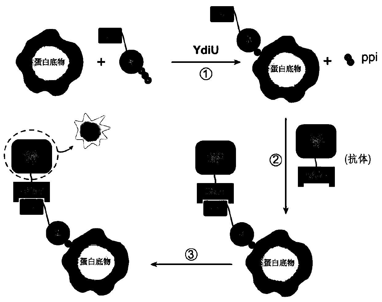
[0025] The present invention comprises the following steps:

[0026] A After adding YdiU protein and substrate to the reaction system, mix well and add 1 μl of Biotin-16-UTP, react at 30-40°C for 1 hour;

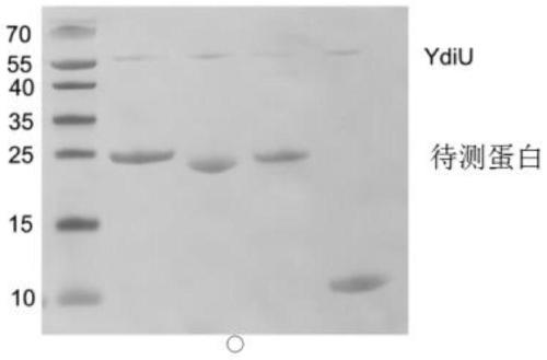
[0027] After the B reaction sample is added to the loading buffer, use 12% SDS-PAGE gel electrophoresis. After electrophoresis, the sample is transferred to the nitrocellulose membrane and then stained with the whole protein of the staining agent;

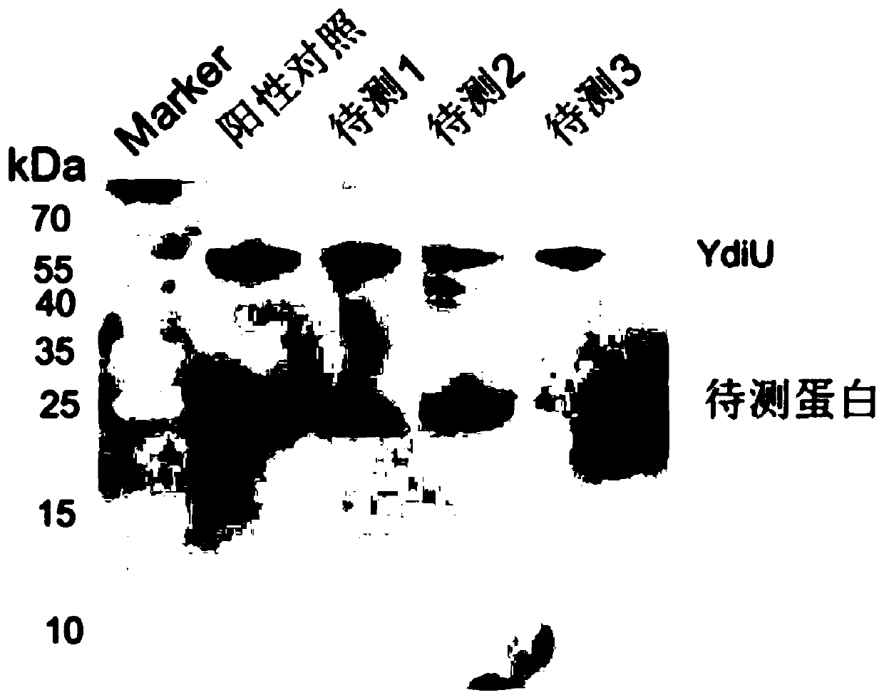
[0028] C. Add the diluted horseradish peroxidase-avidin antibody to the cellulose acetate membrane containing the sample and incubate at room temperature for 30-60 minutes;

[0029] D After washing and blocking for 5min-10min, the membrane can be developed with ECL chromogenic solution. What is visualized is the protein modified by monophosphate u...


PUM


Abstract

The invention discloses a method for detecting uridine monophosphate modification of protein. The method comprises the following steps: by using biotin-labeled UTP (Biotin-16-UTP) as a raw material, under the action of uridine monophosphate transferase YdiU, the protein occurring UMP modification having biotin labeling; and subsequently, verifying whether the protein to be detected has a biotin label or not by using a biotin-streptavidin imprinting method. The method has the characteristics of simplicity, convenience, high sensitivity and the like, can be used for high-throughput screening ofproteins subjected to uridine monophosphate acidification in prokaryotic and eukaryotic cells, can determine modification sites in combination with mass spectrometry detection, and provides an effective identification and research means for research of protein uracil mononucleotide modification.

Description

technical field [0001] The invention relates to the field of biochemistry, in particular to a method for detecting protein monophosphate urylation modification. Background technique [0002] Protein is the bearer of life activities, protein post-translational modification can regulate a variety of life activities, and its imbalance can cause various diseases such as cancer. Among them, protein uracil mononucleotide modification (UMPylation, UMPylation) was first identified in Escherichia coli in 1975, but the substrate protein of UMPylation was limited to the glutamic acid synthase system, resulting in UMPylation has been unknown. It has received attention, although it has been discovered for more than 40 years, the corresponding modification detection method has not been established. With the development of protein modification omics technology, more than 40 UMP-modified proteins have been identified in recent years, indicating that UMP-modified proteins play an important ...

Claims


Application Information

Patent Timeline
IPC IPC(8): G01N21/78G01N1/28G01N1/30G01N33/68
CPCG01N21/78G01N1/30G01N33/6848
Inventor 李冰清杨银龙岳盈盈宋楠楠李翠玲
Owner INST OF BASIC MEDICINE OF SAMS
Why Eureka
Try Eureka