High-specificity exosome separation, detection and enrichment method

A high-specificity exosome technology, applied in cell dissociation methods, biochemical equipment and methods, microbial measurement/testing, etc., can solve problems such as unreported, lack of characterization, and unconvincing test results. Achieve the effects of improved sensitivity, high sensitivity and selectivity, and high in situ amplification efficiency

Active Publication Date: 2020-08-11
SHANGHAI HOSPITAL OF TRADITIONAL CHINESE MEDICINE
View PDF4 Cites 10 Cited by
  • Summary
  • Abstract
  • Description
  • Claims
  • Application Information

AI Technical Summary

Problems solved by technology

[0006] Although these methods have made great progress in the detection of exosomes, there are still deficiencies: 1. Whether it is an immunological detection method based on the antigen-antibody reaction, or the high interaction between aptamers and exosome membrane proteins, The nucleic acid detection method that specifically recognizes and binds may be triggered by free exosomal membrane proteins in t...

Method used


Image

  • High-specificity exosome separation, detection and enrichment method
  • High-specificity exosome separation, detection and enrichment method
  • High-specificity exosome separation, detection and enrichment method

Examples


Embodiment 1

[0049] Embodiment 1 Pre-experiment

[0050] 1 Experimental materials

[0051] Chemicals, Cell Lines and Reagents

[0052] All oligonucleotide sequences in Table 1 were purchased from Shanghai Sangong Bioengineering Technology and Service Co., Ltd. (Shanghai, China). Tris-borate EDTA buffer, DNA staining gel red, 30% polyacrylamide, DPEC water, Dulbecco's PBS, BSA, TMMED and APS were purchased from Shanghai Sanyuan Bioengineering Technology and Service Co., Ltd. (Shanghai, China). T4 DNA ligase, T4 polynucleotide kinase, Phi29 DNA polymerase and RiboLock RNase inhibitor were purchased from Thermo Fisher Scientific Co., Ltd. (Beijing, China). In RPMI1640 medium (Hyclone) containing 10% fetal bovine serum (Hyclone) and 1% penicillin-streptomycin (Hyclone), in 5% CO 2 The A549 cell line was cultured at 37°C under atmosphere. Supernatant cell cultures were obtained from A549 cells. Confocal images were captured by an LSM 780SYSTEM (Zeiss, Germany) and observed using a JEM-1400...

Embodiment 2

[0070] Embodiment 2 system design and operation mechanism

[0071] 1 Experimental method

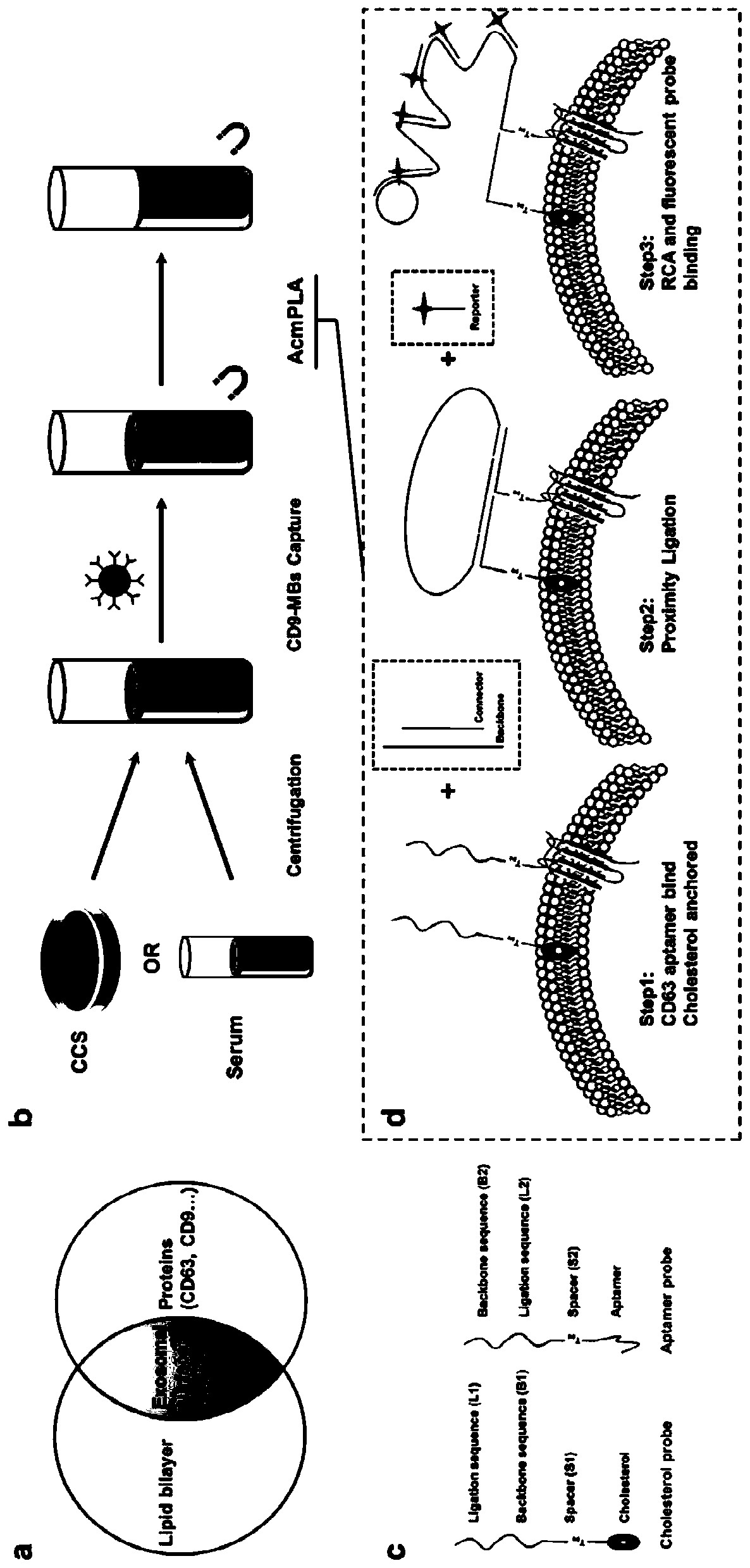
[0072] An aptamer-cholesterol-mediated proximity ligation assay (AcmPLA) was employed to specifically capture exosome surface proteins and minimize nonspecific background signals.

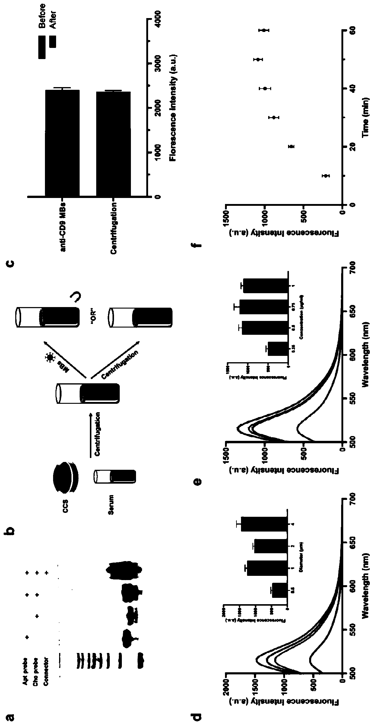
[0073] In the proposed strategy, cell culture supernatant (CCS) or serum samples are centrifuged to remove unwanted particles. The unrefined supernatant obtained consists of exosomes, free proteins and some other biological components. Then, CD9 antibody-labeled magnetic beads (anti-CD9 MBs) were used to capture and assemble CD9-positive biological components under a magnet. To achieve accurate exosome recognition, three active ingredients are used in AcmPLA. The first is two single-stranded DNA (ssDNA) with three functional domains each: a CD63 aptamer probe (Apt probe) and a cholesterol probe (Cho probe). In cholesterol probes, cholesterol that can be inserted into the lipid bilayer is labeled at the 5'...

Embodiment 3

[0077] Example 3 Research on Exosome Extraction and Probe Identification

[0078] 1 Experimental method

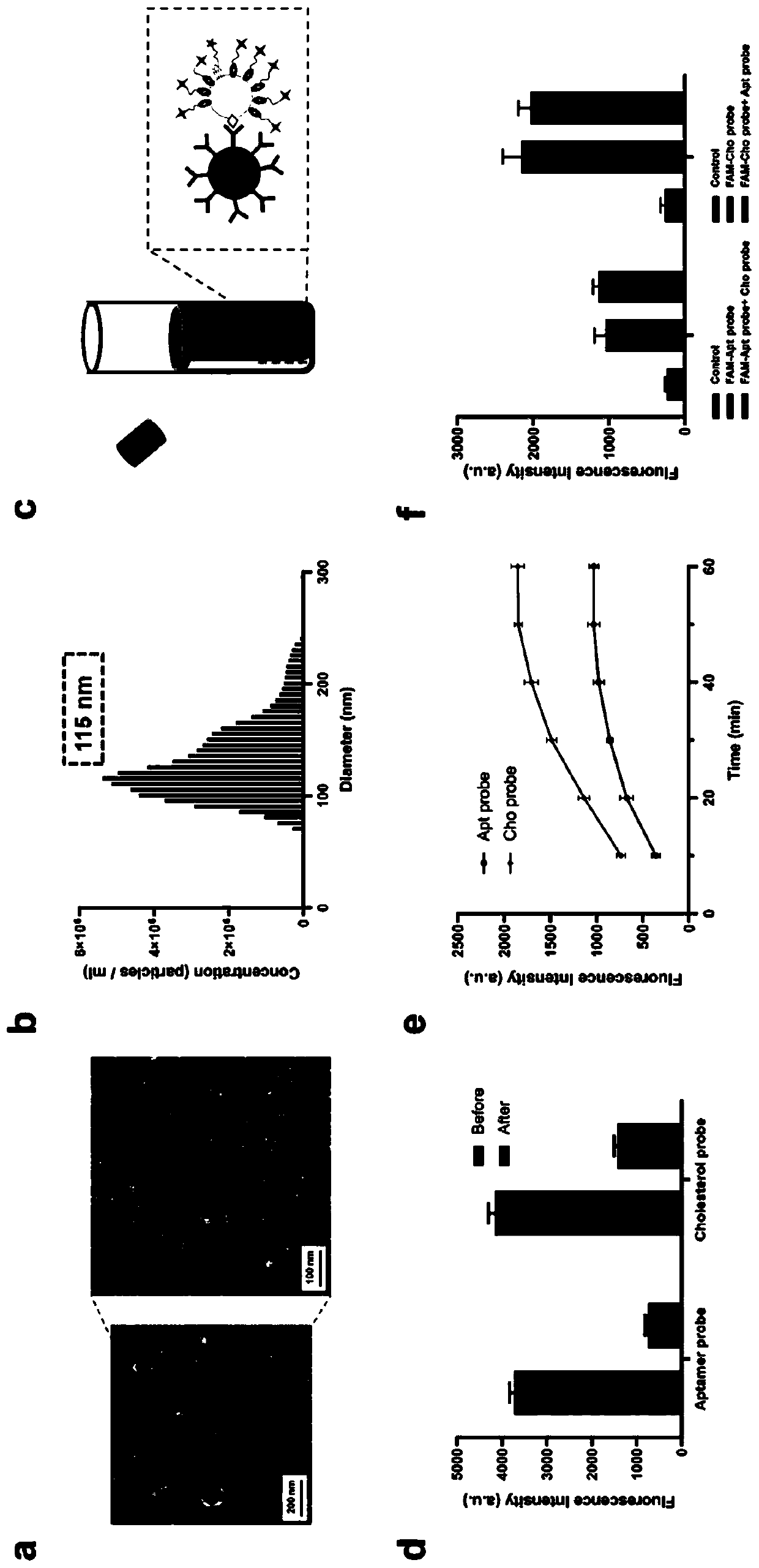
[0079] (1) To investigate whether AcmPLA can be used for accurate exosome identification, we used extracted exosomes obtained from A549 cell CCS by differential centrifugation as a simple model. The isolated exosomes were rigorously characterized using TEM and NTA for morphology observation and protein validation using western blotting. The peak at 115 nm in the diameter distribution determined by NTA is consistent with previous literature. We then detected the isolated exosomes and the proteins (CD9 / CD63 / CD81) expressed on the surface of A549 cells by western blotting.

[0080] (2) Considering that the binding affinity of aptamer probe and cholesterol probe is the key factor to determine the labeling efficiency of AcmPLA, we tested the FAM-labeled CD63 aptamer probe and FAM-labeled cholesterol probe with exosomes, respectively. ( figure 2 c). To avoid undesired fluo...


PUM

PropertyMeasurementUnit
Diameteraaaaaaaaaa

Abstract

The invention relates to the technical field of organism detection and particularly relates to a high-specificity exosome separation, detection and enrichment method which comprises the following steps of constructing a nucleic acid amplification reaction which can be triggered only when phospholipid bilayer and corresponding membrane protein on an exosome membrane exist simultaneously, realizinghigh-specificity exosome detection, and enriching the exosome by virtue of magnetic beads. The method is advantaged in that 1, for a traditional exosome detection method based on the aptamer or the antigen-antibody, in the practical application process, an exosome detection kit is possibly triggered by free exosome membrane proteins such as CD63 protein and CD9 protein in the external environment,a problem that the finally detected signal cannot reflect the quantity of exosomes completely is solved; and 2, the specificity of exosome detection and separation is remarkably improved, interference of free exosome membrane protein in an external environment on exosome detection is reduced, and meanwhile, the higher sensitivity of exosome detection is ensured,

Description

technical field [0001] The invention relates to the technical field of biological detection, in particular to a highly specific exosome isolation, detection and enrichment method. Background technique [0002] Exosomes are vesicles with a uniform size and a diameter of 40-100 nm that are secreted extracellularly after intracellular lysosomal vesicles invaginate to form multi-vesicular bodies (multi-vesicular bodies) that fuse with the cell membrane. Exosomes can carry a variety of proteins, mRNA, miRNA, cholesterol, and sphingomyelin, and participate in processes such as cell communication, cell migration, angiogenesis, and tumor cell growth. Exosomes are an ideal source of potential biomarker molecules because they can reflect the physiological and functional state of their parental cells, are abundant and highly stable in blood, urine and other body fluids, and are easy to obtain samples non-invasively. Because of this, exosome-based detection technology has attracted mor...

Claims


Application Information

Patent Timeline
IPC IPC(8): G01N33/569C12Q1/682C12N15/11G01N1/34C12N5/09
CPCG01N33/56966C12Q1/682G01N1/34C12N5/0693C12N2509/00C12Q2525/205C12Q2563/107
Inventor 毛发江左玲任建琳
Owner SHANGHAI HOSPITAL OF TRADITIONAL CHINESE MEDICINE
Why Eureka
Try Eureka