Method and system for positioning region of interest in real time based on PET data

A region of interest, real-time positioning technology, applied in the field of medical imaging, can solve the problems of prolonging the operation time, the time of sedation or anesthesia for patients, the error of breathing rhythm changes, and the limited help of tumor positioning, so as to simplify the interventional treatment process and eliminate respiratory movement The effect of reducing acquisition time and anesthesia time

Active Publication Date: 2020-08-18
赛诺联合医疗科技(北京)有限公司
View PDF2 Cites 4 Cited by
  • Summary
  • Abstract
  • Description
  • Claims
  • Application Information

AI Technical Summary

Problems solved by technology

However, in actual operation, even if the patient has been trained, the change of breathing rhythm will still inevitably bring about errors.
In addition, PET acquisition during consecutive respiratory cycles may prolong the duration of surgery and patient sedation or anesthesia
More importantly, this method is of limited help for real-time tumor localization for thermal ablation (radiofrequency and microwave)

Method used


Image

  • Method and system for positioning region of interest in real time based on PET data
  • Method and system for positioning region of interest in real time based on PET data
  • Method and system for positioning region of interest in real time based on PET data

Examples


Embodiment 1

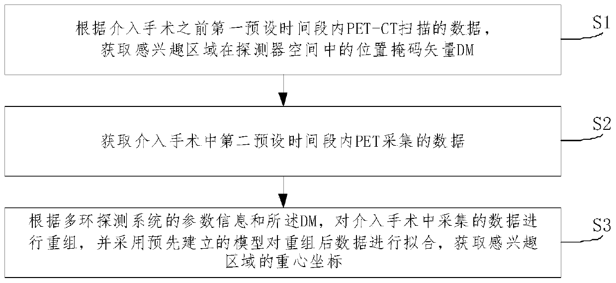
[0056] Such as figure 1 As shown, the embodiment of the present invention provides a method for real-time positioning of a region of interest based on PET data. The execution subject can be a workstation / computing device of a multi-ring detection system, which specifically includes the following steps:

[0057] Step S1, according to the PET-CT scan data within the first preset time period before the interventional operation, obtain the position mask vector DM of the region of interest in the detector space.

[0058] In step S1, taking PET-CT as an example, the coordinates of the region of interest are obtained by relying on PET acquisition data. The same method is also applicable to other matching modality scanning systems, such as PET-MR.

[0059] Step S2, obtaining the data collected by PET within the second preset time period in the interventional operation;

[0060] It should be noted that in this step, the artificially set parameter information (acquisition parameters an...

Embodiment 2

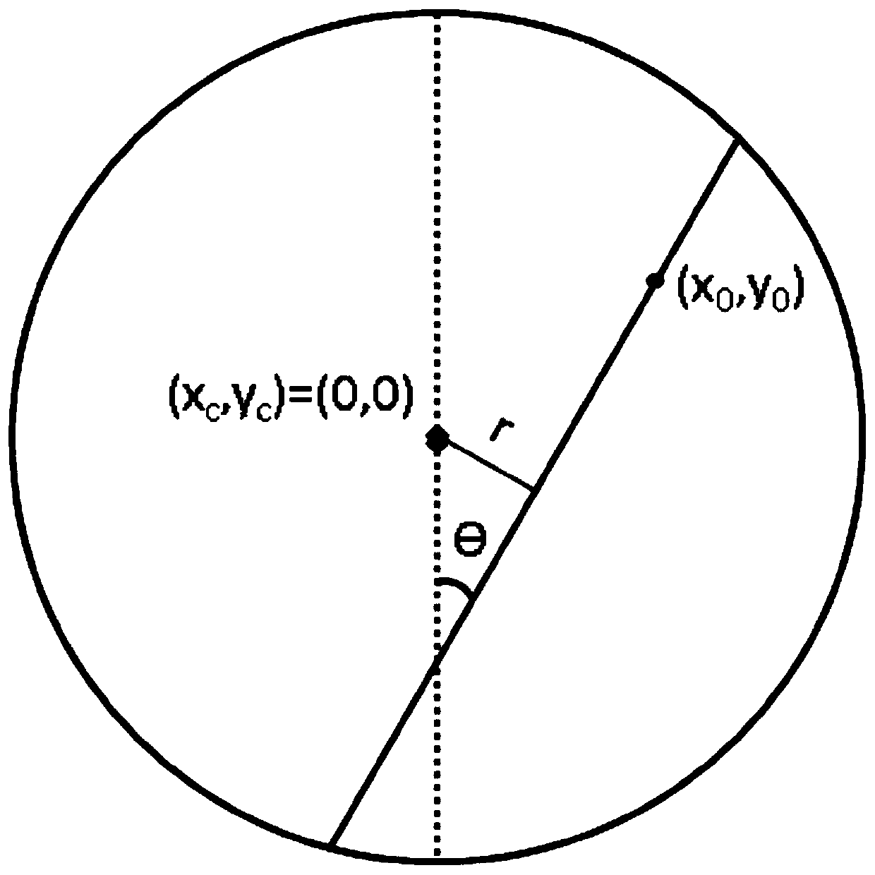
[0067] combine Figure 2 to Figure 5 As shown, the embodiment of the present invention provides a method for real-time positioning of a region of interest based on PET data. The execution subject can be a workstation / computing device of a multi-ring detection system, and the region of interest can be a tumor region. In this embodiment, the tumor location The real-time positioning method is not limited to periodic changes affected by respiration, of course, it is also applicable to other situations, such as heartbeat, accidental movement of patients and so on.

[0068] In this embodiment, short-term PET imaging is used to delineate the tumor area requiring interventional therapy, and then the tumor trajectory is fitted in the detector sinogram space, and the three-dimensional space coordinates of the delineated tumor are calculated in real time. Taking PET-CT scanning as an example, the specific steps of this method are as follows:

[0069] Step A1: Before the interventional o...


PUM


Abstract

The invention discloses a PET data-based region of interest real-time positioning method and system. The method comprises the following steps: S1, obtaining a position mask vector DM of a region of interest in a detector space according to PET-CT scanning data in a first preset time period before an interventional operation; S2, acquiring data acquired by PET in a second preset time period in theinterventional operation; and S3, according to the parameter information of the multi-ring detection system and the DM, recombining the data collected in the interventional operation, and processing the recombined data by adopting a pre-established model to obtain the barycentric coordinates of the region of interest. The method is beneficial to application of a PET gated imaging technology, motion artifacts are reduced, and the image quality is improved; the position of interest influenced by respiratory movement can be determined in real time, and the movement influence is eliminated.

Description

technical field [0001] The invention relates to the field of medical imaging, in particular to a method and system for real-time positioning of a region of interest based on PET data that can be used in a positron emission computed tomography imaging system. Background technique [0002] Positron Emission Tomography PET (Positron Emission Tomography) is a high-end nuclear medicine imaging diagnostic equipment. In practice the use of radionuclides (eg 18 F. 11 C, etc.) Label the metabolites and inject the nuclides into the human body, and then perform functional metabolic imaging on the patient through the PET system to reflect the situation of life metabolic activities. PET scans are usually accompanied by other modality scans (such as CT, MRI, etc.) to obtain imaging of the patient's anatomy. The two images will eventually be fused on the same machine, compatible with the advantages of functional imaging and anatomical imaging, to achieve the purpose of early detection o...

Claims


Application Information

Patent Timeline
Patent Type & Authority Applications(China)
IPC IPC(8): A61B6/03A61B6/12
CPCA61B6/037A61B6/469A61B6/12Y02A90/10
Inventor 李楠崔洁
Owner 赛诺联合医疗科技(北京)有限公司
Why Eureka
Try Eureka