System and method for evaluating joint pressure by multiple-modality imaging

A multi-modal imaging and joint technology, applied in medical science, sensors, diagnostic recording/measurement, etc., can solve the problems of image blur, high price, low time resolution, etc., and achieve the effect of eliminating image blur and eliminating errors

Pending Publication Date: 2020-09-18
连云港市第二人民医院
View PDF4 Cites 1 Cited by
  • Summary
  • Abstract
  • Description
  • Claims
  • Application Information

AI Technical Summary

Problems solved by technology

[0003] The methods used to evaluate joint diseases in the prior art include MRI, CT, and X-rays to obtain images of joints and their soft tissues. However, both CT and X-rays use the grayscale of X-rays to represent soft tissues or bones. For differences in X-ray absorption, its spatial resolution is low and it is not sensitive enough to diseased tissues; while MRI imaging has high spatial resolution, its time resolution is not high, and it is poor for structures with low proton density, and it also involves The use of magnetic contrast agents is expensive, and it is not easy to apply to the prognosis assessment of joint diseases
[0004] At the same time, there is no tool that can quantify the intra-articular pressure in the clinical medical process. If the pressure in the ...

Method used


Image

  • System and method for evaluating joint pressure by multiple-modality imaging
  • System and method for evaluating joint pressure by multiple-modality imaging
  • System and method for evaluating joint pressure by multiple-modality imaging

Examples


Embodiment 1

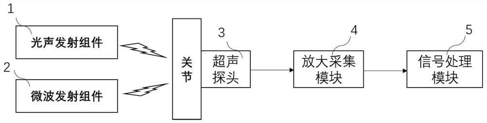
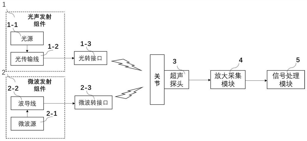
[0037] see Figure 1-2 , a multi-modal imaging system for evaluating joint pressure, comprising a photoacoustic emission assembly 1, a microwave emission assembly 2, an ultrasound probe 3, an amplification acquisition module 4, and a signal processing module 5; the photoacoustic emission assembly 1 and the microwave emission assembly 2. Alternately transmit light signals and microwave signals to the joints, the ultrasonic probe 3 is used to detect the photoacoustic signals and microwave thermoacoustic signals generated at the joints; the amplification acquisition module 4 is used to amplify and collect the photoacoustic signals signal and microwave thermoacoustic signal, and transmitted to the signal processing module 5; the signal processing module 5 generates a photoacoustic image and a thermoacoustic image from the photoacoustic signal and microwave thermoacoustic signal, and converts the photoacoustic image and thermal Acoustic image superposition reconstruction into multi...

Embodiment 2

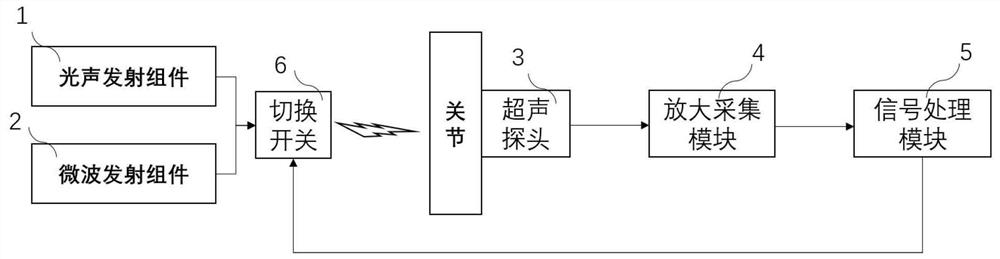
[0047] Refer to attached Figure 3-4 , a multi-modal imaging system for evaluating joint pressure, comprising a photoacoustic emission assembly 1, a microwave emission assembly 2, an ultrasound probe 3, an amplification acquisition module 4, and a signal processing module 5; the photoacoustic emission assembly 1 and the microwave emission assembly 2 The switch 6 controlled by the signal processing module 5 alternately transmits optical signals and microwave signals to the joints, and the ultrasonic probe 3 is used to detect the photoacoustic signals and microwave thermoacoustic signals generated at the joints; the amplification acquisition module 4 is used to amplify and collect the photoacoustic signal and microwave thermoacoustic signal, and transmit it to the signal processing module 5; the signal processing module 5 generates a photoacoustic image and a thermoacoustic image from the photoacoustic signal and microwave thermoacoustic signal , and superpose and reconstruct th...

Embodiment 3

[0058] In order to further illustrate how to use multi-modal images to better realize the assessment of joint pressure, the present invention also discloses a method for evaluating joint pressure with multi-modal imaging, the steps are as follows:

[0059] Step 1. Acquiring thermoacoustic signals and photoacoustic signals at the joints to be evaluated;

[0060] Step 2. Reconstructing a thermoacoustic image by using a filter back projection algorithm according to the thermoacoustic signal, and reconstructing a photoacoustic image by using a delayed superposition algorithm according to the photoacoustic signal;

[0061] Step 3: Analyzing multiple joint points on the thermoacoustic image as feature points, finding corresponding multiple feature points in the photoacoustic image for registration, and using an image fusion algorithm to obtain the fused multiple joint points. modal image;

[0062] Step 4. Obtain thermoacoustic signals and photoacoustic signals at joints to be evalu...


PUM


Abstract

The invention discloses a system for evaluating joint pressure by multiple-modality imaging. The system is characterized in that the system comprises an acousto-optic emission component, a microwave emission component, an ultrasonic probe, an amplification and collection module and a signal processing module, wherein the acousto-optic emission component and the microwave emission component alternately emit a light signal and a microwave signal to a joint; the ultrasonic probe is used for detecting an acousto-optic signal and a microwave thermo-acoustic signal generated on the joint; the amplification and collection module is used for amplifying and collecting the acousto-optic signal and the microwave thermo-acoustic signal, and transmitting the acousto-optic signal and the microwave thermo-acoustic signal to the signal processing module; and the signal processing module generates an acousto-optic image and a thermo-acoustic image by the acousto-optic signal and the microwave thermo-acoustic signal, and overlaps and reconstructs the acousto-optic image and the thermo-acoustic image to obtain a multiple-modality image. According to the system, an influence on joint evaluation by fuzzy microwave thermal imaging can be eliminated, and more accurate information in the joint, including local pressure, abrasion and the like, in the joint can be obtained.

Description

technical field [0001] The invention relates to the technical field of joint disease prognosis assessment, in particular to a multimodal imaging system and method for assessing joint pressure. Background technique [0002] Abnormal changes in intra-articular pressure are the cause of many diseases. Take knee osteoarthritis as an example. Congenital or acquired reasons lead to increased local pressure on the knee joint, which leads to excessive wear in daily activities, which leads to cartilage wear and tear. Osteoarthritis develops. [0003] The methods used to evaluate joint diseases in the prior art include MRI, CT, and X-rays to obtain images of joints and their soft tissues. However, both CT and X-rays use the grayscale of X-rays to represent soft tissues or bones. For differences in X-ray absorption, its spatial resolution is low and it is not sensitive enough to diseased tissues; while MRI imaging has high spatial resolution, its time resolution is not high, and it is...

Claims


Application Information

Patent Timeline
IPC IPC(8): A61B5/00A61B5/05
CPCA61B5/0035A61B5/0095A61B5/0507A61B5/4528
Inventor 刘明明李琳迪吕南宁李梦迪许海燕程建华臻贾学军张浩
Owner 连云港市第二人民医院
Why Eureka
Try Eureka