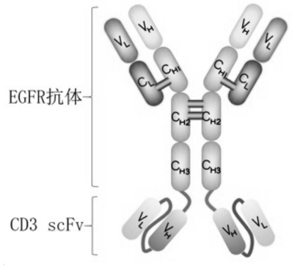
EGFR-CD3 bifunctional antibody and application thereof
An EGFR-CD3, bifunctional antibody technology, applied in applications, antibodies, anti-tumor drugs, etc., can solve the problems of increasing the risk of nervous system complications, host rejection, etc., to promote differentiation and proliferation, and reduce immunogenicity. Effect
- Summary
- Abstract
- Description
- Claims
- Application Information
AI Technical Summary
Problems solved by technology
Method used
Image

Examples
Embodiment 1
[0035] Example 1: Antibody Preparation of EGFR-CD3 Bifunctional Antibody
[0036] According to the amino acid sequence matching of the heavy chain and light chain of the EGFR-CD3 bifunctional antibody, the heavy chain Cetuxi-H1L1, Cetuxi-H2L2, Cetuxi-H3L3, Cetuxi-H4L1, Cetuxi-H2L1, and Cetuxi-H3L2 of the bifunctional antibody were artificially synthesized, respectively. and light chain Cetuxi-L cDNA and constructed into pcDNA3.1 vector. The light chain plasmid and the heavy chain plasmid of the bifunctional antibody identified by sequencing were co-transfected into 293F cells at a weight ratio of 1:1.
[0037] Electroporation conditions: 4mm click cup, voltage 250v, 5mm, cells 1×10 8 . The electroporated cells were placed at 37° C., 5% CO2, and cultured at 130 rpm / min for 7 days. After the cells were cultured for 7 days, the supernatant was collected by centrifugation. The supernatant was centrifuged at 6000 rpm for 10 min, and the final concentration of 200 mM NaCl and 50 ...
Embodiment 2
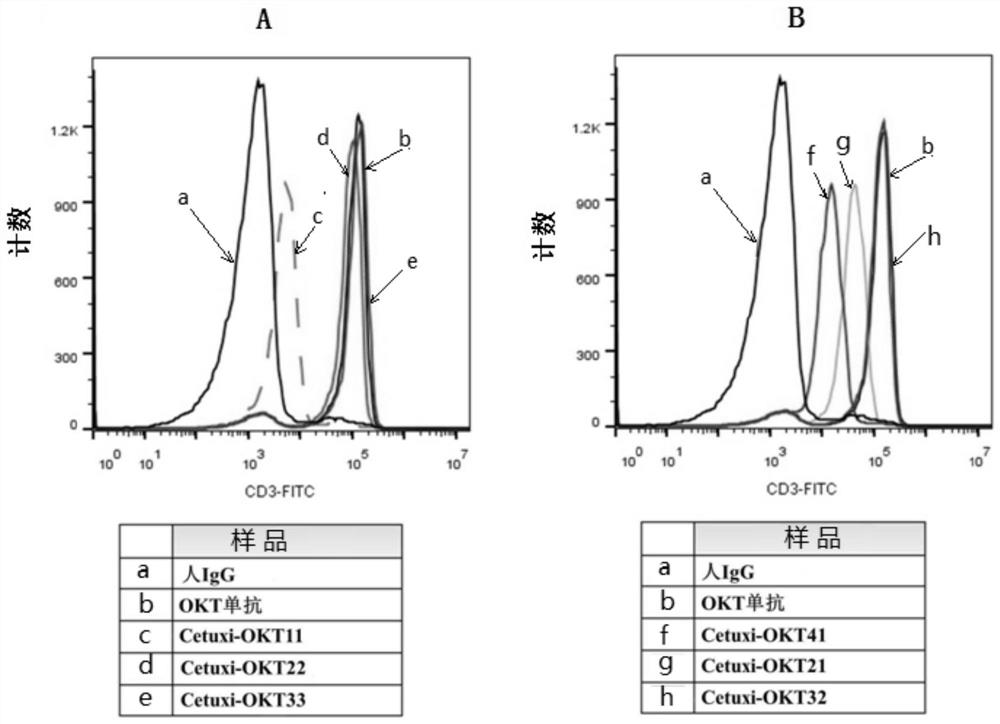
[0038] Example 2: Detection of binding activity of EGFR-CD3 bifunctional antibody to CD3 protein on the surface of Jurkat cells
[0039] The cell surface protein binding ability of each EGFR-CD3 bifunctional antibody (Cetuxi-OKT11, Cetuxi-OKT22, Cetuxi-OKT33, Cetuxi-OKT41, Cetuxi-OKT21 and Cetuxi-OKT32) was detected by flow cytometry. The binding ability of EGFR-CD3 bifunctional antibody to CD3 protein on the surface of Jurkat cells was detected. The negative control of the experiment was set up as human IgG antibody, the positive control was humanized OKT clone antibody (produced by Sino Biological), and the experimental group was each expressed and purified EGFR-CD3 bifunctional antibody. Set detection cells to 1×10 6 Each tube was added with 2 μg / ml of each antibody and incubated on ice for 30 min. After washing the cells by centrifugation, Alexa 488 fluorescein-labeled mouse anti-human secondary antibody (diluted 1:200) was added and incubated on ice for 30 min. After t...
Embodiment 3
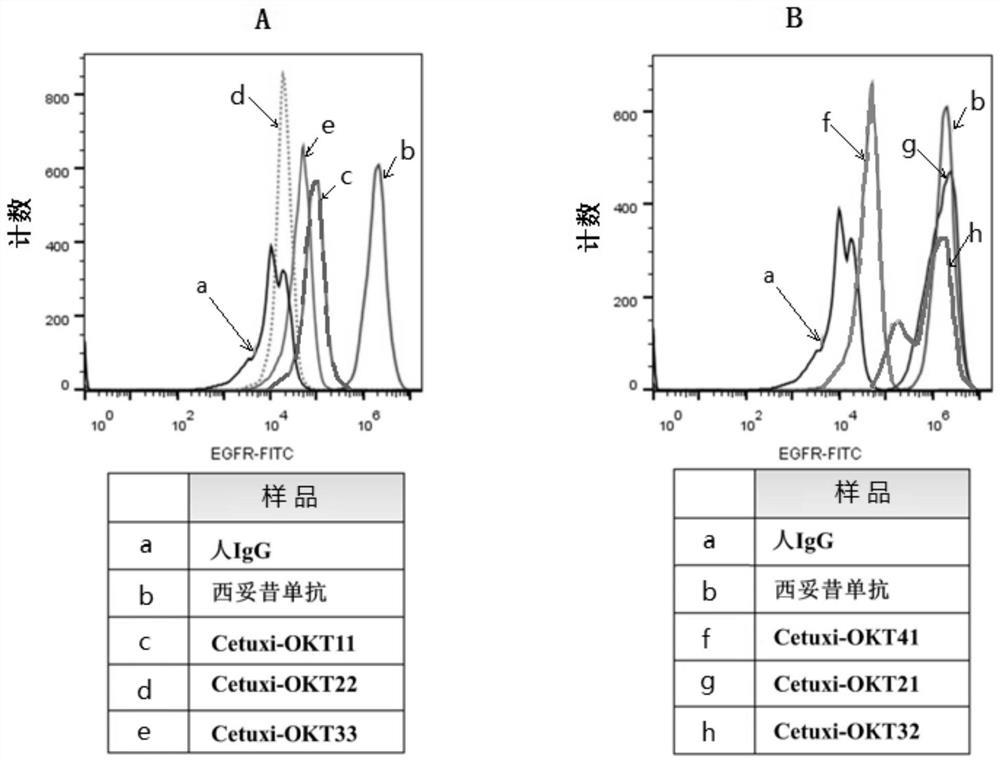
[0041] Example 3: EGFR-CD3 bifunctional antibody detection of EGFR protein binding activity on the surface of epithelial tumor cell A431 cells
[0042] The cell surface protein binding ability of each bifunctional antibody was detected by flow cytometry. The binding ability of the bifunctional antibody to the EGFR protein on the surface of A431 cells was detected. The negative control of the experiment was set as human IgG antibody, the positive control was human-mouse chimeric antibody (Erbitux monoclonal antibody, namely cetuximab), and the experimental group was each expressed and purified bifunctional antibody. Set detection cells to 1×10 6 Each tube was added with 2 μg / ml of each antibody and incubated on ice for 30 min. After washing the cells by centrifugation, Alexa488 fluorescein-labeled mouse anti-human secondary antibody (1:200 dilution) was added and incubated on ice for 30 min. After the incubation is complete, wash the cells by centrifugation. Fluorescent lab...
PUM
 Abstract
Description
Claims
Application Information