Choroid three-dimensional blood vessel imaging and quantitative analysis method and device based on optical coherence tomography system

An optical coherence tomography and scanning system technology, applied in the field of choroidal three-dimensional vascular imaging and quantitative analysis, can solve the problems of poor signal-to-noise ratio, difficult automatic segmentation, and low contrast of blood vessels

Pending Publication Date: 2020-10-30
WENZHOU MEDICAL UNIV
View PDF9 Cites 7 Cited by
  • Summary
  • Abstract
  • Description
  • Claims
  • Application Information

AI Technical Summary

Problems solved by technology

However, the current system does not solve the problem of poor signal-to-noise ratio of deep choroidal vessels caused by backscattering attenuation, which leads to difficulties in vessel boundary segmentation
For this reason, most of the current quantitative choroidal vessel analysis algorithms in OCT images only use traditional image processing methods. However, since the choroid is located below the RPE, the light passes through the RPE and is attenuated by pigment absorption. At the same time, the choroid itself is rich in pigment. The attenuation of the light signal occurs in the depth of the choroid, resulting in blurred boundaries between the choroid and the pigment epithelium and between the choroid and the sclera
In addition, due to the existence of vascular (low signal) and non-vascular (high signal) components in the choroid, the contrast with the inner and outer boundaries is low, and the boundary continuity is poor, so it is difficult to realize the automatic segmentation of the boundary with the traditional segmentation method including the shortest path graph theory algorithm
Therefore, the accuracy of automatic differentiation is low, and manual correction after a large number of boundary detections is required, which is time-consuming
Because the brightness of the choroidal signal in the OCT image is affected by related factors such as the RPE layer, the type of OCT instrument, and the focus during operation, the separation of choroidal vessels and stroma based on the fixed threshold method has great limitations in theory. Poor adaptability

Method used


Image

  • Choroid three-dimensional blood vessel imaging and quantitative analysis method and device based on optical coherence tomography system
  • Choroid three-dimensional blood vessel imaging and quantitative analysis method and device based on optical coherence tomography system
  • Choroid three-dimensional blood vessel imaging and quantitative analysis method and device based on optical coherence tomography system

Examples


Embodiment Construction

[0099] The present invention can be better described below in conjunction with the accompanying drawings and some specific embodiments.

[0100] (1) Data acquisition and preprocessing:

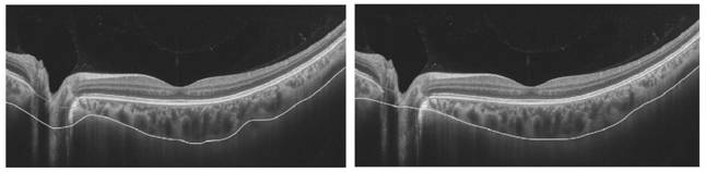
[0101] Use the current commercial equipment or self-built OCT to obtain fundus images, preprocess the images including appropriate cropping, and retain the OCT choroidal intensity map, as shown in the attached figure 2 shown.

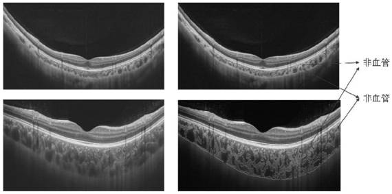
[0102] (2) Reverse attenuation compensation for choroidal signal:

[0103]The signals received by the OCT detector are backscattered and reflected signals. Due to the influence of RPE and choroid’s own pigments on light absorption, light scattering of a certain wavelength is lost. By extracting the principle and law of choroid scattering light attenuation and constructing a signal compensation and enhancement algorithm, the visualization and contrast of choroid images can be improved.

[0104] The attenuation correction processing algorithm of the OCT signal include...


PUM


Abstract

The invention discloses a choroid three-dimensional blood vessel imaging and quantitative analysis method and device based on an optical coherence tomography system. Reverse compensation is performedon a deep choroid blood vessel and a matrix, which are obtained by OCT, through a backward scattering attenuation signal based on the idea of OCT backward scattering signal reverse compensation, and the signal-to-noise ratio of the choroid blood vessel is improved; after an enhanced OCT choroid image signal is acquired, a segmentation method based on deep learning is further adopted to perform intelligent segmentation on boundaries between a choroid and a retina epithelium and between the choroid and a sclera, and the defects that a traditional algorithm consumes time and is low in accuracy inthree-dimensional choroid boundary segmentation are overcome; and, on the basis of boundary segmentation, an improved self-adaptive threshold segmentation method is further adopted to automatically separate three-dimensional choroidal vessels from non-vascular tissues, and global and regional quantitative indexes capable of representing choroidal ischemia are calculated according to the distribution and proportion of the vessels in the image in a three-dimensional body space.

Description

technical field [0001] The invention relates to the technical field of OCT, in particular to a method and device for three-dimensional choroidal vascular imaging and quantitative analysis based on an optical coherence tomography system. Background technique [0002] The choroid is a highly vascularized, pigment-rich tissue located between the retina and sclera. The main function is to provide oxygen and nutrients to the RPE and the outer layer of the retina. The oxygen it transports accounts for about 90% of the oxygen consumption of the retina, which is necessary to maintain the high metabolic activity of the photoreceptor cells in the outer layer of the retina; especially in the fovea without blood vessels area, the choroid is the only way for its material exchange. Many diseases are closely related to abnormal vascular structure of the choroid, such as age-related maculopathy, high myopia maculopathy, diabetic retinopathy and so on. Therefore, it is of great significanc...

Claims


Application Information

Patent Timeline
Patent Type & Authority Applications(China)
IPC IPC(8): G06T7/11G06T7/12G06T7/136G06T11/00G06N3/04G06N3/08A61B3/10A61B3/12
CPCG06T7/11G06T7/12G06T7/136G06T11/008G06N3/08A61B3/102A61B3/1225G06T2207/10101G06T2207/30101G06T2207/30041G06T2207/20056G06T2207/20081G06T2207/20084G06N3/045
Inventor 周翔天沈梅晓吴昊陈思思金梓
Owner WENZHOU MEDICAL UNIV
Why Eureka
Try Eureka