Atomic force microscope colloid probe surface modification method

An atomic force microscope, surface modification technology, applied in scanning probe technology, scanning probe microscopy, measuring devices, etc. Large roughness and other problems, to achieve high universality, improve yield and mass production efficiency, and achieve a wide range of effects

Inactive Publication Date: 2020-12-15
KUNMING UNIV OF SCI & TECH
View PDF1 Cites 3 Cited by
  • Summary
  • Abstract
  • Description
  • Claims
  • Application Information

AI Technical Summary

Problems solved by technology

However, often because the added adhesive such as FeOOH is adsorbed on the surface of the colloid, it cannot be evenly distributed evenly, so even the colloidal microspheres with single-layer functional group ligands adsorbed by electrostatic interaction and conforming to the langmuir adsorption model often have a large particle size. roughness
Especially for colloidal microspheres with unimolecular adsorption, extreme roughness will affect the accuracy and reliability of the measurement of the interaction force

Method used


Image

  • Atomic force microscope colloid probe surface modification method
  • Atomic force microscope colloid probe surface modification method
  • Atomic force microscope colloid probe surface modification method

Examples


Embodiment 1

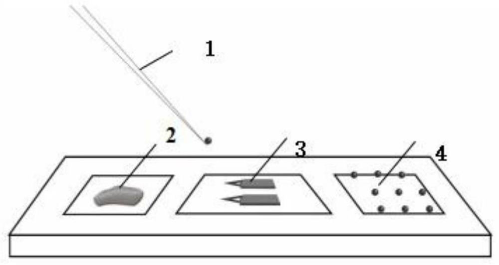
[0032] A method for surface modification of an atomic force microscope colloidal probe, comprising the steps of:

[0033] 1) Preparation of colloidal microspheres wrapped in hematite film: at room temperature, slowly to 0.01M FeCl 3 A minimum amount of 1M NaOH solution was added to the solution to pH7. will be 5×10 7 / ml 5μm silica microspheres were added to 1mL FeOOH solution and ultrasonicated for 10min. Put the centrifuge tube containing the sample solution into a centrifuge and centrifuge at a centrifugal speed of 5000 rpm for 5 minutes. After repeated washing with ultrapure water for 3 times, the colloidal microspheres loaded with iron oxyhydroxide were placed in an oven at a temperature of 100 degrees Celsius and dried for 12 hours.

[0034] 2) Place the colloidal microspheres wrapped with the hematite nanofilm in the tannic acid modification solution, and shake slowly for 24 hours. The centrifuge tube containing the sample solution was put into a centrifuge and cent...

Embodiment 2

[0042] A colloidal microsphere probe modification method for an atomic force microscope, comprising the steps of:

[0043] 1) Preparation of colloidal microspheres wrapped in hematite film: at room temperature, slowly to 0.01M FeCl 3 A minimum amount of 1M NaOH solution was added to the solution to pH7. will be 5×10 7 / ml 2μm polyethylene terephthalate microspheres were added to 0.5mL FeOOH solution and ultrasonicated for 20min. Put the centrifuge tube containing the sample solution into a centrifuge and centrifuge at a centrifugal speed of 5000 rpm for 10 minutes. After repeated washing with ultrapure water for 3 times, the colloidal microspheres loaded with iron oxyhydroxide were placed in an oven at a temperature of 100 degrees Celsius and dried for 12 hours.

[0044] 2) Place the colloidal microspheres wrapped with the hematite nanofilm in the humic acid modification solution, and shake slowly for 48 hours. The centrifuge tube containing the sample solution was put int...

Embodiment 3

[0053] A colloidal microsphere probe modification method for an atomic force microscope, comprising the steps of:

[0054] 1) Preparation of colloidal microspheres wrapped in hematite film: at room temperature, slowly to 0.01M FeCl 3 A minimum amount of 1M NaOH solution was added to the solution to pH7. will be 5×10 7 / ml of glass microspheres with a diameter of 10 μm was added to 2 mL of FeOOH solution and ultrasonicated for 30 min. Put the centrifuge tube containing the sample solution into a centrifuge and centrifuge at a centrifugal speed of 5000 rpm for 5 minutes. After repeated washing with ultrapure water for 3 times, the colloidal microspheres loaded with iron oxyhydroxide were placed in an oven at a temperature of 100 degrees Celsius and dried for 12 hours.

[0055] 2) Place the colloidal microspheres wrapped with the hematite nanofilm in the fulvic acid modification solution, and shake slowly for 24 hours. The centrifuge tube containing the sample solution was pu...


PUM

PropertyMeasurementUnit
diameteraaaaaaaaaa

Abstract

The invention discloses an atomic force microscope colloid probe surface modification method, and belongs to the technical field of atomic force microscope probe modification. The invention provides ahigh-success-rate colloid probe synthesis and surface modification method, and the interaction force between colloid and minerals can be measured more accurately. The method comprises the following steps: immersing colloidal microspheres in a FeOOH solution for a period of time, taking out the colloidal microspheres, drying, putting the colloidal microspheres in a functional group modification solution for a certain period of time, and carrying out cold drying; dipping a needle head of a 30G injector in a minimum amount of epoxy resin mixed liquid, transferring the epoxy resin mixed liquid tothe front end of a cantilever of the probe, sucking the modified colloidal microspheres by using a new needle head, transferring the colloidal microspheres to the cantilever, and curing epoxy resin to obtain the functional group modified colloidal probe. According to the invention, the yield of the colloid probe can be greatly improved, the surface roughness of the functional group modified probeis reduced, and the experimental error is greatly reduced.

Description

technical field [0001] The invention relates to the technical field of atomic force microscope probe modification, in particular to a method for surface modification of an atomic force microscope colloidal probe. Background technique [0002] AFM is a tool that can measure the morphology of samples under different environmental conditions such as air, vacuum, and liquid. In addition, various surface forces can be qualitatively and quantitatively measured through probe modification, which greatly promotes the measurement technology of intermolecular force and provides an effective method for the measurement of the interaction force between functional groups. This can not only observe the energy required to break a single molecular bond, but also observe the interaction force between macroscopic colloidal particles. However, because ordinary needle-point probes are often not accurate enough to detect some small interactions, certain errors occur. Therefore, the magnitude of ...

Claims


Application Information

Patent Timeline
Patent Type & Authority Applications(China)
IPC IPC(8): G01Q60/38
CPCG01Q60/38
Inventor 王振全赛克·高希张迪董蕙苏博何南
Owner KUNMING UNIV OF SCI & TECH
Why Eureka
Try Eureka