
Skin cancer classification method based on optical intensity and gradient of OCT imaging image

A classification method, skin cancer technology, applied in the field of biomedical image processing

Pending Publication Date: 2020-12-22
NINGBO UNIVERSITY OF TECHNOLOGY
View PDF0 Cites 1 Cited by
  • Summary
  • Abstract
  • Description
  • Claims
  • Application Information

AI Technical Summary

Problems solved by technology

However, there may be agreement between intensity-based and gradient-based features, which may reveal common patterns for classifying diseased skin tissue

Method used


Image

  • Skin cancer classification method based on optical intensity and gradient of OCT imaging image
  • Skin cancer classification method based on optical intensity and gradient of OCT imaging image
  • Skin cancer classification method based on optical intensity and gradient of OCT imaging image

Examples


Embodiment Construction

[0042] The present invention will be described in detail below in conjunction with the accompanying drawings and specific embodiments.

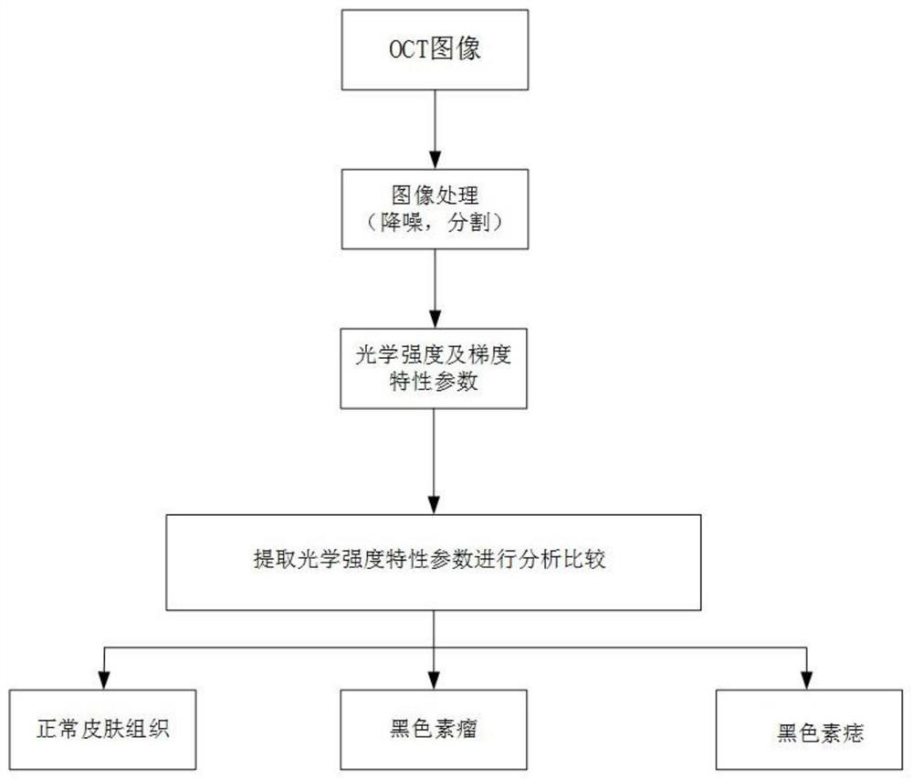
[0043] A skin cancer classification method based on optical intensity and gradient of OCT imaging images, the method comprising:

[0044] First, the intensities in the OCT images are normalized to [0, 1].

[0045] Second, background noise was removed from all OCT images. Background noise is any light other than the light being monitored. Types of background noise include ambient light (such as sun rays) and lighting in a laboratory. In the field of image noise removal, preventing or reducing background noise is important. In this patent, a simple method is used to remove background noise. We select a small region of fixed size in each OCT image at the top edge of each OCT image. The average intensity value in that region is then calculated and treated as background noise. Then, a noise-filtered OCT image was obtained by subtracting the ...


PUM


Abstract

The invention discloses a skin cancer classification method based on the optical intensity and gradient of an OCT imaging image, and the method comprises the steps: (1) collecting a skin cancer imagethrough an OCT device, and normalizing the intensity in the OCT image into [0, 1]; (2) performing noise reduction on the OCT image through noise reduction processing; (3) extracting optical intensitycharacteristic parameters; (4) calculating to obtain the discrete value of each parameter of the marked sample, and then obtaining the difference and ROC analysis of each characteristic parameter in the marked control group through a mathematical statistics method to obtain a boundary value; and (5) calculating to obtain the discrete value of each parameter of the unknown sample, comparing the discrete value with the boundary value in the step (4), and classifying the unknown sample according to the comparison result. According to the invention, normal skin tissues, melanoma and melanin nevuscan be quickly and accurately judged; discomfort and trauma brought to a patient by an invasive method of skin biopsy are avoided.

Description

technical field [0001] The invention relates to a skin cancer classification method based on the optical intensity and gradient of an optical coherence tomography (OCT) imaging image, belonging to the field of biomedical image processing. Background technique [0002] Skin cancer is one of the most common cancers worldwide. According to the World Health Organization, more than 2 million cases of skin cancer are diagnosed each year. In clinical practice, skin biopsy is the gold standard for diagnosing skin cancer. However, skin biopsy is an invasive procedure that can cause patient discomfort. Therefore, non-invasive methods have been adopted to eliminate the shortcomings of biopsy in skin cancer diagnosis, including dermoscopy, confocal microscopy, Raman spectroscopy and optical coherence tomography (OCT). These patterns are able to generate images to help dermatologists diagnose different types of skin disorders. Dermoscopy not only allows a visual inspection of the ski...

Claims


Application Information

Patent Timeline
IPC IPC(8): G06K9/62G06T5/00
CPCG06T2207/10101G06T2207/30088G06T2207/20032G06T2207/30096G06F18/24G06T5/70
Inventor 高巍
Owner NINGBO UNIVERSITY OF TECHNOLOGY
Learn More