

Rich double-staining kit and use method thereof
A technology of dyeing reagents and kits, which is applied in the preparation of test samples, biological testing, sampling, etc., and can solve the problem of the difficulty in accurately distinguishing the positional relationship between tumor cells and elastic fibers, the difficulty in interpreting the pleura of lung cancer, and the difficulty in accurately displaying the relationship between tumor cells and the pleura. Elastic fiber layer relationship and other issues
- Summary
- Abstract
- Description
- Claims
- Application Information
AI Technical Summary
Problems solved by technology
Method used
Image

Examples
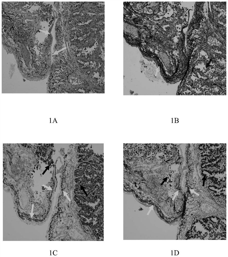
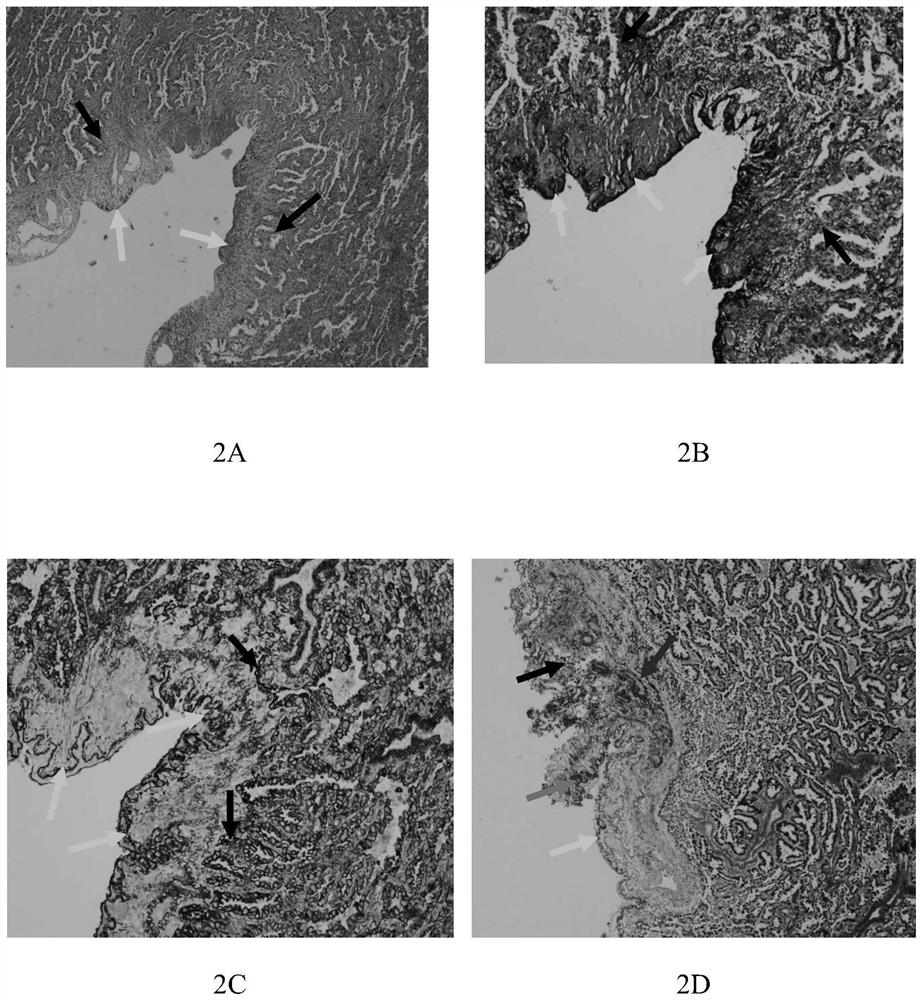
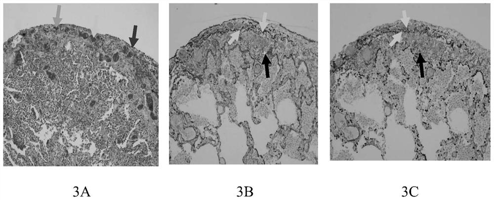
Embodiment 1
[0067] Example 1 Ordinary elastic fiber dyeing method
[0068] In this embodiment, the elastic fiber dyeing kit purchased from Yili Company was used, referring to the instruction manual, and carried out according to the following operations:
[0069] 1) Paraffin tissue with conventional standardized treatment, sliced at 3-5 μm;
[0070] 2) Bake in a 70°C incubator for 2 hours;
[0071] 3) Conventional xylene dewaxing twice, 10min each time, and then hydrating with gradient ethanol for a few seconds with 100% ethanol twice, 95% ethanol aqueous solution twice, 85% ethanol aqueous solution once, fully rinsed with tap water, and rinsed with distilled water 3 times;
[0072] 4) Add iron iodine hematoxylin for 10-15 minutes, wash with water;
[0073] 5) Differentiate the differentiation solution for a few seconds until the excess liquid fades away;
[0074] 6) Wash with water, add 95% ethanol to remove iodine for 1min;
[0075] 7) Counterstain in VG solution for 1 min;
[0...
Embodiment 2
[0080] Example 2 Double staining method using anti-TTF-1 antibody as primary antibody of the present invention
[0081] For paraffin tissue with routine standardized treatment, select the tissue block that needs to be stained, slice 3-5 μm, and then bake in a 70°C incubator for 2 hours; conventional xylene dewaxing 2 times, 10 minutes each time; then 100% ethanol 2 times each time 3 minutes each time, 95% ethanol water solution twice for 3 minutes each time, 85% ethanol water solution once for 3 minutes each time, followed by gradient ethanol hydration for 10 seconds, fully rinsed with tap water, rinsed with distilled water for 3 times; put the slices into pH 9.0, 1mM EDTA antigen retrieval solution Heated in a pressure cooker to 100°C for 1-5 minutes (2-3 minutes after gassing), cooled naturally to room temperature, washed 3 times with distilled water, 3 minutes each time; then sliced in 3% hydrogen peroxide solution at room temperature Incubate for 10 minutes to remove e...
Embodiment 3
[0085] Example 3 Double staining method using AE1 / AE3 as the primary antibody of the present invention
[0086] For routine normalized paraffin tissue, select the tissue block that needs to be stained, slice 3-5 μm, and then bake in a 70°C incubator for 2 hours; conventional xylene dewaxing twice, each 10min; and then in 100% ethanol twice 3 minutes each time, 95% ethanol water solution twice for 3 minutes each time, 85% ethanol water solution once for 3 minutes each time, followed by gradient ethanol hydration for 10 seconds, fully rinsed with tap water, rinsed with distilled water for 3 times; put the slices into pH 9.0, 1mM EDTA antigen retrieval solution Heat it in a pressure cooker to 100°C for 1-5min (continue 2-3min after gassing), cool naturally to room temperature, wash 3 times with distilled water, 3min each time; then place the slices in 3% aqueous hydrogen peroxide solution, Incubate at room temperature for 10 minutes to remove endogenous peroxidase, then wash wi...
PUM
| Property | Measurement | Unit |
|---|---|---|
| thickness | aaaaa | aaaaa |
Abstract
Description
Claims
Application Information