Method for preparing bionic hollow human body part model through 3D printing

A human body part, 3D printing technology, applied in processing and manufacturing, liquid material additive processing, household components, etc., can solve the problems of long model time, high cost, stitching, repeated tearing, etc.

Pending Publication Date: 2021-03-16
NAT INST CORP OF ADDITIVE MFG XIAN
View PDF8 Cites 3 Cited by
  • Summary
  • Abstract
  • Description
  • Claims
  • Application Information

AI Technical Summary

Problems solved by technology

[0005] In order to solve the shortcomings of the traditional casting and injection molding process, such as long time to prepare models, high cost, small individual differences, and 3D printed medical models cannot be used in surgical suturing, repeated tearing and other training applications, the present invention provides a 3D printing preparation bionic hollow human body Part Model Method

Method used


Image

  • Method for preparing bionic hollow human body part model through 3D printing
  • Method for preparing bionic hollow human body part model through 3D printing
  • Method for preparing bionic hollow human body part model through 3D printing

Examples


Embodiment 1

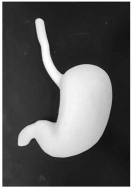
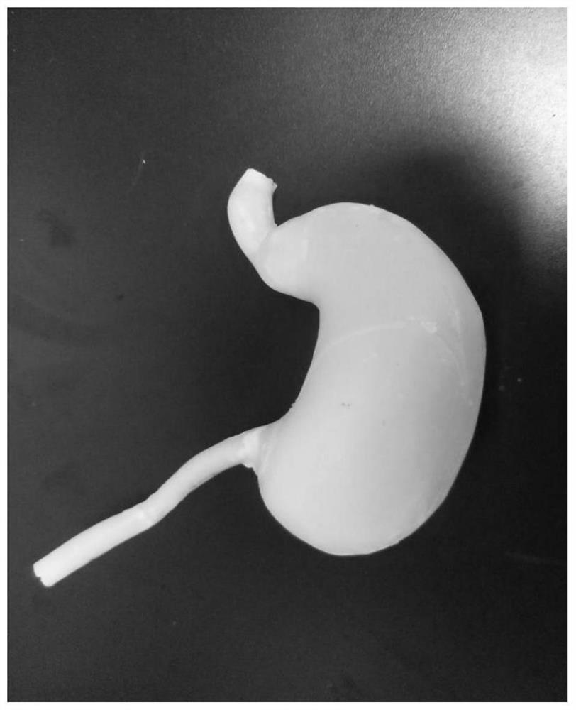
[0098] Such as figure 1 As shown, this embodiment realizes the preparation of the bionic hollow human stomach and esophagus model through the following steps:

[0099] 1. Data acquisition and extraction:

[0100] Scan the human stomach and esophagus model by scanning equipment (3D laser scanner) to obtain its point cloud data;

[0101] Because the model data in this embodiment is obtained from the existing gastroesophageal model, it is not necessary to obtain data from special medical images.

[0102] 2. Data processing and mold design:

[0103] Use software (Geomagics Studio) to process the point cloud data of the human gastroesophagus, making it a data format recognizable by a 3D printer that meets the requirements.

[0104] 3. Build a model:

[0105] Professionally painted Figure three Dimensional software (Solidworks, ProE, UG, Fluent) special modules, using the data obtained in step 2, to establish the shell mold model, hollow inner mold model and assembly mold mode...

Embodiment 2

[0121] This embodiment realizes the preparation of the bionic hollow human stomach and esophagus model through the following steps:

[0122] 1. Data acquisition and extraction:

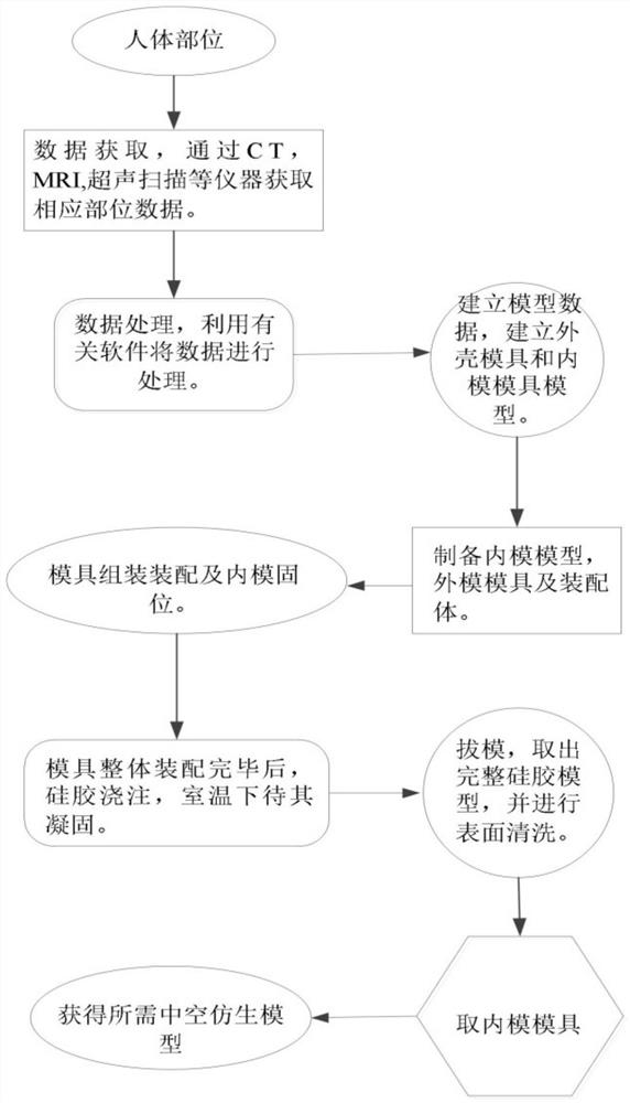
[0123] Use medical imaging equipment, such as CT, MRI, PET-CT and ultrasound scanners, to detect the human gastroesophagus, obtain and save the corresponding image data, and extract and record the surface elastic modulus of the gastroesophagus;

[0124] 2. Data processing:

[0125] Import CT, MRI, PET-CT image data into Mimics, complete segmentation processing, and then repair the image data after segmentation processing by 3-matic and Magics, and then detect it by Geomagic data model to make it become three-dimensional data, which meets the requirements A data format that a 3D printer can recognize.

[0126] 3. Building a model

[0127] Professionally painted Figure three Dimensional software (Solidworks, ProE, UG, Fluent) special modules use the data obtained in step 2 to establish the shell mo...

Embodiment 3

[0141] This embodiment realizes the preparation of the bionic hollow human stomach and esophagus model through the following steps:

[0142] 1. Obtain and extract data:

[0143] Use medical imaging equipment, such as CT, MRI, PET-CT and ultrasound scanners, to detect the human gastroesophagus, obtain and save the corresponding image data, and extract and record the surface elastic modulus of the gastroesophagus;

[0144] 2. Data processing:

[0145] Use medical software (Mimics, 3-matic, Magics, Geomagics) to process the human gastroesophageal image data obtained in step 1, and convert it into a data format that can be recognized by 3D printing equipment;

[0146] The specific processing process is: import CT, MRI, PET-CT, and image data into Mimics, complete the extraction and segmentation processing, and then repair the image data after segmentation processing by 3-matic and Magics, and then detect it by the Geomagic data model to make it a Three-dimensional data becomes a...


PUM


Abstract

The invention relates to a method for preparing a human body part model, in particular to a method for preparing a bionic hollow human body part model through 3D printing. The defects that a traditional casting and injection molding technology is long in model preparation time, high in cost and small in individual difference, and a medical model obtained through 3D printing cannot be applied to training such as surgical suture and repeated tearing are overcome. The method comprises the steps of data acquisition, data processing, model building, shell mold, hollow inner mold and assembly part mold preparation, mold treatment, mold assembly and retention, silica gel infusion, mold drawing and the like. The bionic hollow human body part model prepared through the method is real and is close to the real surface hardness of a target human body part, the surface is smooth and not sticky, the thickness and distribution of the model are consistent with those of the human body part, the tissuestructure is observed obviously, and repeated tearing is resisted. The method is suitable for surgical simulation, suture surgery training, clinical teaching and the like.

Description

technical field [0001] The invention relates to a method for preparing a human body part model, in particular to a method for preparing a bionic hollow human body part model by 3D printing. Background technique [0002] Gastroesophageal reflux disease is a disease in which gastric content reflux causes esophageal symptoms and complications. Clinically, it mainly manifests as heartburn, acid reflux, and substernal pain. Esophageal cancer, etc.; its etiology is relatively complicated, and it is currently considered to be an upper gastrointestinal motility disorder. Surgical treatment is considered to be the best way for gastroesophageal diseases. Laparoscopic fundoplication does not require resection and no sampling is required. Reconstruction is required, the image is magnified, the light is good, the operation can be performed in a narrow space, the trauma is small, and the recovery is fast. It has become one of the main ways to treat the disease, and the rational choice of ...

Claims


Application Information

Patent Timeline
Patent Type & Authority Applications(China)
IPC IPC(8): B29C64/106B33Y10/00B29K83/00
CPCB29C64/106B33Y10/00B29K2083/005B29L2031/7028
Inventor 王影卢秉恒王晶王远
Owner NAT INST CORP OF ADDITIVE MFG XIAN
Why Eureka
Try Eureka