Pig stomach bottom gland stem cell separation and three-dimensional organ culture method

A culture method and organoid technology, which is applied in the field of porcine fundic gland stem cell separation and three-dimensional organoid culture, can solve the problems of inapplicability of large economic animals, achieve good organoid formation rate and survival rate, and increase organoid formation rate effect on survival

Pending Publication Date: 2021-04-06
INST OF SUBTROPICAL AGRI CHINESE ACAD OF SCI
View PDF2 Cites 0 Cited by
  • Summary
  • Abstract
  • Description
  • Claims
  • Application Information

AI Technical Summary

Problems solved by technology

However, in the field of animal nutrition research, the method of establishing organoids based on the physiological and biochemical characteristics of mouse and human stomach is not suitable for large economic animals such as pigs

Method used

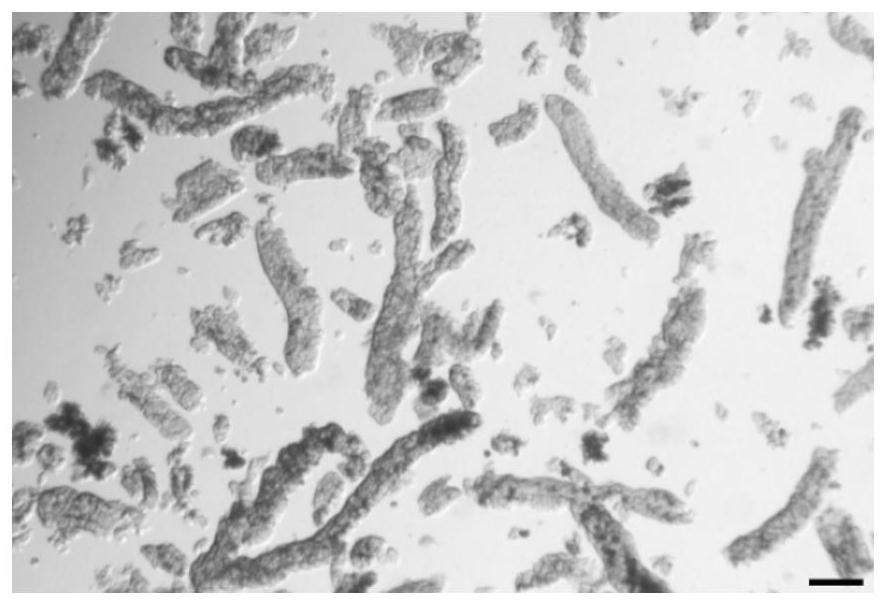
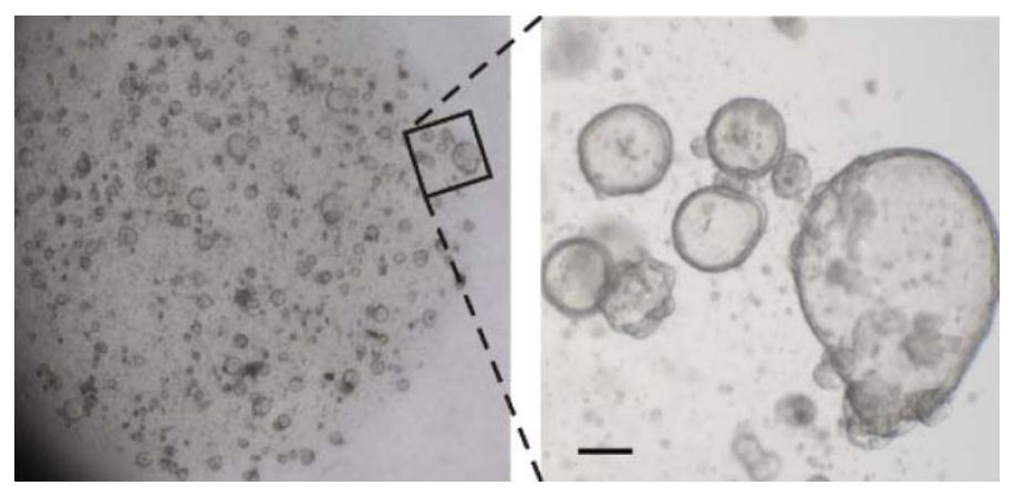
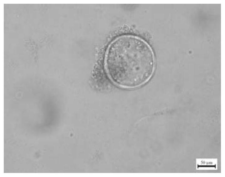

Image

  • Pig stomach bottom gland stem cell separation and three-dimensional organ culture method
  • Pig stomach bottom gland stem cell separation and three-dimensional organ culture method
  • Pig stomach bottom gland stem cell separation and three-dimensional organ culture method

Examples


Embodiment 1

[0050] Isolation of porcine fundic gland stem cells and three-dimensional organoid culture method:

[0051] A. Isolation of Fundic Gland Stem Cells:

[0052] (1) Before the start of the experiment, sterilize consumables such as scissors and tweezers that have been steam sterilized and dried with 100% alcohol, put the 24-well cell culture plate in a 37°C incubator to preheat for at least 30 minutes, and thaw Matrigel on ice.

[0053] (2) Preparation of flushing solution: Prepare 5× mother solution in 100 mL and store at 4°C. The specific ingredients are shown in Table 1:

[0054] Table 1

[0055]

[0056] Immediately before the separation of gastric basilar gland tissue, at room temperature (15-25°C), add sterilized water to the 5× mother solution and dilute it to 1× working solution, and then add 0.5mM, DL-dithiothreitol (dithiothreitol sugar alcohol), fully mixed, that is, the flushing solution, which needs to be prepared and used immediately.

[0057] (3) The followin...


PUM


Abstract

The invention discloses a pig stomach bottom gland stem cell separation and three-dimensional organoid culture method. The method comprises three parts of dissociation and separation of stomach bottom gland stem cells, preparation of a pig stomach organoid culture medium and three-dimensional stomach organoid culture. According to the invention, the blank of a stomach organ technology in pig nutrition research is filled, and a new tool and a carrier are provided for the stomach nutrition function research of monogastric animals.

Description

technical field [0001] The invention belongs to the technical field of cell culture, in particular to a method for isolating porcine fundic gland stem cells and culturing three-dimensional organoids. Background technique [0002] The core issue of animal nutrition research is how to improve the conversion rate of dietary protein to animal protein, and the stomach is the main organ in the digestive system responsible for the initial processing and decomposition of the diet, and the study of its nutritional function is of great significance for improving feed utilization. For monogastric animals such as pigs, gastric divisions mainly include pyloric gland area, fundic gland area, aglandular area, and cardia gland area. The tissue structure of the stomach is divided into four layers, which are the mucosal layer, submucosa layer, muscular layer, and serosa layer from the cavity to the outside. The innermost layer of the mucosal layer is a single-layer columnar epithelium, which...

Claims


Application Information

Patent Timeline
IPC IPC(8): C12N5/074
CPCC12N5/068C12N2509/00C12N2509/10C12N2513/00C12N2500/32C12N2501/11C12N2501/119C12N2501/15C12N2500/38C12N2501/345C12N2500/46C12N2501/415C12N2533/90C12N2501/405
Inventor 周健熊霞万丹邵宜锐吴宇梁印遇龙
Owner INST OF SUBTROPICAL AGRI CHINESE ACAD OF SCI
Why Eureka
Try Eureka