Colorectal cancer detection kit
A kit and quantitative detection technology, applied in the field of immunoassay, to achieve the effect of simple microplate reader, fast and sensitive detection, simple and easy operation
- Summary
- Abstract
- Description
- Claims
- Application Information
AI Technical Summary
Problems solved by technology
Method used
Image

Examples
Embodiment 1
[0022] Example 1. Preparation and purification of anti-CEA antibody
[0023] CHO cells stably expressing CEA were cultured with DMEM medium, and 10 7 CHO-CEA + The cells were injected intraperitoneally into female Balb / c mice together with Freund's complete adjuvant for initial immunization, followed by 10 injections every 2 weeks 7 CHO-CEA + The cells were injected intraperitoneally into the above-mentioned female Balb / c mice, and the immunization was continued. Finally, the last immunization was carried out 3 days before the fusion with myeloma cells, and the final immunization was carried out with 10 7 CHO-CEA + Cells were immunized intravenously. Get immunized Balb / c mouse splenocytes, fuse it with myeloma Sp2 / 0 cell line, and use Iscove medium (0.1 mM hypoxanthine, 0.4 μ M aminopterin) to contain 10% serum after fusion and 16 μM thymidine), diluted to an appropriate concentration, added to a 96-well plate for cultivation. After 10 days, the cell supernatant was tak...
Embodiment 2
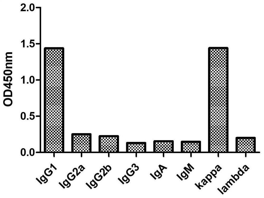
[0025] Example 2. Anti-CEA antibody subtype identification
[0026] The subtype of the anti-CEA monoclonal antibody Cab-207 obtained in Example 1 was identified by ELISA (the kit was purchased from Proteintech). The ELISA plate provided in the kit has been pre-coated with specific antibodies against mouse IgG1, IgG2a, IgG2b, IgG3, IgA, IgM, kappa light chain, lambda light chain, and the anti-CEA purified in Example 1 Antibody sample Cab-207 was added to the sample well, 50 μl per well, without incubation. Add 1X goat anti-mouse IgA+IgM+IgG-HRP into the sample wells, 50 μl per well, mix gently, and incubate for 1 h. Remove the liquid in the well and add 1XPBST to wash the well 3 times, and absorb excess water with absorbent paper. Add chromogenic solution, 100 μl per well, and develop color for 15 minutes at room temperature in the dark. Add 100 μl stop solution to stop the color reaction. Detect the OD value at 450nm by microplate reader, the result is as follows figure 1...
Embodiment 3
[0027] Example 3. Monoclonal Antibody Sequence Determination
[0028] After recovering the hybridoma cell line corresponding to Cab-207, culture it to a total number of 10 7 Cells were collected by centrifugation at 1000rpm for 5min, and RNA was extracted. Add TRNzol-A+ to the cell pellet for lysis, and let it stand at room temperature for 15 minutes. Add 200μl chloroform per ml TRNzol-A+, vortex for 15 seconds, and let stand for 3 minutes. Centrifuge at 13000rpm at 4°C for 10 minutes. Trizol-A+ cell solution is divided into three layers: the upper colorless aqueous phase, the middle layer and the lower yellow organic phase. Transfer the aqueous phase containing RNA to a centrifuge tube, and add an equal volume of isopropanol, mix well, and let stand at room temperature for 25 minutes. Centrifuge at 13,000 rpm at 4°C for 10 minutes, discard the waste liquid to obtain the RNA pellet that sinks to the bottom. After the RNA pellet was washed twice with 75% ethanol, the RNA wa...
PUM
 Abstract
Description
Claims
Application Information
 - R&D Engineer
- R&D Manager
- IP Professional
- Industry Leading Data Capabilities
- Powerful AI technology
- Patent DNA Extraction
Browse by: Latest US Patents, China's latest patents, Technical Efficacy Thesaurus, Application Domain, Technology Topic.
© 2024 PatSnap. All rights reserved.Legal|Privacy policy|Modern Slavery Act Transparency Statement|Sitemap