
Method for increasing tissue exosome yield by using freezing microtome

A technology of frozen sections and exosomes, applied in tissue culture, cell dissociation methods, preparation of test samples, etc., can solve the problems of long time-consuming, cumbersome process, and increased extraction yield, so as to increase the tissue surface area and reduce the dosage , The effect of increasing production

Active Publication Date: 2021-06-25
北京恩泽康泰生物科技有限公司
View PDF8 Cites 0 Cited by
  • Summary
  • Abstract
  • Description
  • Claims
  • Application Information

AI Technical Summary

Problems solved by technology

Existing methods for extracting tissue exosomes generally use steps such as shearing, enzymatic hydrolysis, centrifugation, ultracentrifugation, and ultrafiltration. For example, Chinese patent CN107523536A discloses an extraction method and application of tissue-derived exosomes. The core Extract tissue exosomes based on mechanical shearing, enzymatic lysis of tissue, overnight sedimentation with PEG, centrifugation and other steps, which takes a long time, and there may be a large amount of protein impurities in the extracted exosomes; Chinese patent CN111621471A discloses a soft tissue cell The extraction method and application of extracellular vesicles, extracting soft tissue exosomes through steps such as shearing, centrifugation, and ultrafiltration, is also a cumbersome and time-consuming process; Chinese patent 2020115224931.3 discloses a method for rapidly extracting tissue extracellular vesicles Separation and enrichment method, through enzymatic hydrolysis, differential centrifugation, ultracentrifugation, molecular exclusion, and ultrafiltration, the rapid extraction of tissue exosomes is realized
Most of the existing patents and documents optimize the methods of enzymatic hydrolysis, centrifugation, and ultrafiltration for tissue exosome extraction to improve the purity and yield of tissue exosome extraction, but there are few methods for tissue exosome extraction. The method is optimized so that tissue exosomes can be extracted quickly and the yield of extraction can be increased

Method used


Image

  • Method for increasing tissue exosome yield by using freezing microtome
  • Method for increasing tissue exosome yield by using freezing microtome
  • Method for increasing tissue exosome yield by using freezing microtome

Examples


Embodiment 1

[0044] Example 1 A method of using a frozen microtome to increase the output of exosomes in mouse brain tissue

[0045] A method for extracting mouse brain tissue exosomes using a frozen microtome, the specific steps are as follows:

[0046] (1) Pretreatment before the experiment: Start the cryostat 3 hours in advance to pre-cool, set the temperature of the microtome and the sample head to -25°C, tissue sample 0.2g, embedding box (Nantong Meiweide Experimental Equipment Co., Ltd., article number EM05 -0707) and the sample tray were pre-cooled in dry ice 30 minutes in advance, and the embedding medium was 1×PBS buffer solution and pre-cooled at 4°C 3 hours in advance.

[0047] (2) Tissue sample embedding: The following operations are carried out in dry ice: put the tissue sample into the embedding box and use pre-cooled 1×PBS buffer solution for embedding, so that the 1×PBS buffer solution is completely submerged in the tissue After the tissue is embedded in PBS, apply a layer...

Embodiment 2

[0069] Example 2 A method of using a frozen microtome to increase the production of exosomes in mouse spleen tissue

[0070] This example provides a method for extracting exosomes from mouse spleen tissue using a cryostat, and the specific steps are as follows:

[0071] (1) Pretreatment before the experiment: start the cryostat 3 hours in advance to pre-cool, set the temperature of the microtome and the sample head to -20°C, pre-cool the tissue samples, embedding boxes and sample trays in dry ice 30 minutes in advance, and pre-cool the embedding agent 3 hours in advance Pre-cool at 4°C.

[0072](2) Tissue sample embedding: Put the tissue sample into the embedding box and use pre-cooled 1×PBS buffer for embedding, so that the 1×PBS buffer is completely submerged in the tissue, and wait for the 1×PBS buffer to wrap. After burying the tissue, apply a layer of 1×PBS buffer solution on the sample tray with a thickness of about 3mm, and quickly transfer the tissue embedded in the e...

Embodiment 3

[0090] Example 3 A method of using a frozen microtome to increase the yield of exosomes in mouse gastric tissue

[0091] This example provides a method for extracting mouse gastric tissue exosomes using a cryostat, the specific steps are as follows:

[0092] (1) Pretreatment before the experiment: start the cryostat 3 hours in advance to pre-cool, set the temperature of the microtome and the sample head to -20°C, pre-cool the tissue samples, embedding boxes and sample trays in dry ice 30 minutes in advance, and pre-cool the embedding agent 3 hours in advance. h Pre-cool at 4°C.

[0093] (2) Tissue sample embedding: Put the tissue sample into the embedding box and use pre-cooled 1×PBS buffer for embedding, so that the 1×PBS buffer is completely submerged in the tissue, and wait for the 1×PBS buffer to wrap. After burying the tissue, apply a layer of 1×PBS buffer on the sample tray with a thickness of about 2mm. When the 1×PBS buffer on the tray turns white, quickly transfer th...


PUM


Abstract

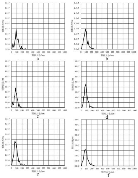
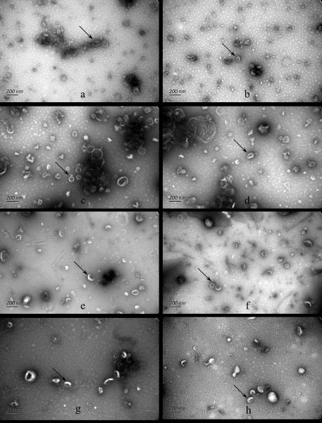
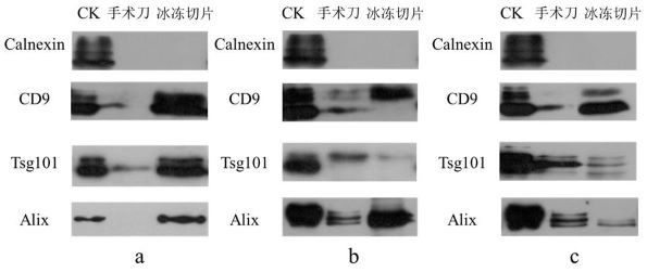
The invention relates to a method for improving the yield of tissue exosomes by using a freezing microtome, and belongs to the technical field of tissue slice preparation and exosome extraction. The method comprises the steps of embedding a tissue sample, slicing the tissue sample by using the freezing microtome, collecting the sample slices, performing enzymolysis, performing ultracentrifugation , performing exclusion, performing ultrafiltration and the like. By adopting the method, the yield of the extracted exosomes is obviously higher than that of the exosomes extracted by other tissue exosome extraction methods in the prior art, the yield of the exosomes of tissue samples with the same dosage is increased by about 2-10 times, the dosage of the tissues is greatly reduced, the problem that enough exosomes are difficult to obtain from various scarce tissue samples can be solved, 0.1-0.2 g of tissue can meet subsequent NTA, WB, electron microscope, transcriptome and other detection and analysis, the practicability is good, and the application prospect is wide.

Description

technical field [0001] The invention relates to the technical field of tissue slice preparation and exosome extraction, in particular to a method for increasing tissue exosome production by using a frozen microtome. Background technique [0002] Exosomes are lipid bilayer-coated particles with a size of 30–150 nm released by different cells. They can wrap and deliver molecules such as RNA, DNA, and protein, and participate in various physiological and pathological pathways as messengers for intercellular communication. At present, the methods for isolating exosomes from body fluids such as blood, urine, and saliva are very mature, but the relevant research and methods for effectively isolating exosomes from the intercellular space of various tissues are still relatively limited. The key link in the process of tissue exosome enrichment is the process of obtaining cell suspension by mechanical shearing, which will cause damage to the cell structure to a certain extent. The ru...

Claims


Application Information

Patent Timeline
IPC IPC(8): G01N1/28G01N1/34G01N1/36G01N1/40G01N1/42C12N5/071C12N5/078C12N5/079C12N5/09
CPCC12N5/0618C12N5/0631C12N5/0648C12N5/0679C12N5/0693C12N2509/00C12N2509/10G01N1/28G01N1/286G01N1/34G01N1/36G01N1/40G01N1/4044G01N1/42G01N2001/2873G01N2001/4088
Inventor 王程程秦伟伟杨瑞程敏林海军孔关义
Owner 北京恩泽康泰生物科技有限公司
Learn More