Method for carrying out lysosome classification by taking lysosome metabolite as marker
A technology for lysosomes and metabolites, applied in the field of lysosome classification, can solve the problem of poor fineness of lysosome classification
- Summary
- Abstract
- Description
- Claims
- Application Information
AI Technical Summary
Problems solved by technology
Method used
Image
Examples
Embodiment 1
[0049] This embodiment takes isolated HEK-293T cells as an example for introduction, and of course the present invention can also be used for mouse embryonic fibroblasts (MEF), mouse lung fibroblasts (MLF), bladder cancer cells (T24), human Immortalized bladder epithelial cells (SV-HUC-1), BxPC-3 cells, mouse cardiomyocytes and cardiac fibroblasts, mouse cerebral cortex neurons and glial cells, mouse peritoneal macrophages, mouse skin Cells such as fibroblasts (MEFs).
[0050] This embodiment discloses a method for classifying lysosomes using lysosomal metabolites as markers, comprising the following steps:
[0051] Step 1. Cell culture
[0052] Culture of HEK-293T cells
[0053] HEK-293T cells used for passage were cultured in T25 culture flasks. Aspirate the culture medium in the culture bottle, add 1 mL of preheated trypsin to wash it again, add 1 mL of trypsin, and put it in a 37°C carbon dioxide incubator to digest for 2 minutes; add 4 mL of preheated culture medium to...
Embodiment 2
[0107] Example of tip (electrode tip) and shape
[0108] Such as Figure 2-5 As shown, among them, figure 2 The tip opening diameters of each glass electrode in Tip1-Tip5 are 272nm, 340nm, 298nm, 288nm, 310nm respectively, and the tip length of the glass electrode is selected as 8mm.
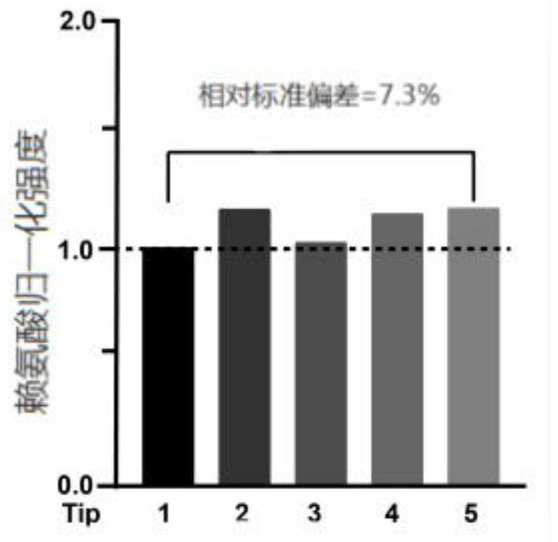
[0109] Reproducibility of SLMS using glass electrodes with similar tip size and shape. figure 2 Scanning electron microscope images of five selected glass pipette tips for the geometrical properties of the SLMS glass electrodes. Scale bar 200 μm. image 3 SLMS measures lysine (LYS, relative standard deviation = 7.3%), Figure 4 Histidine (HIS, relative standard deviation = 8.1%) and Figure 5 Reproducibility of arginine (ARG, relative standard deviation = 7.5%).
[0110] Repeated measurements were made at the levels of 5 ppm (34 μM) of LYS, 5 ppm (32 μM) of HIS and 5 ppm (28 μM) of ARG added to the artificial simulated lysosomal fluid (ALF). ALF contains 145mM NaCl, 5mM KCl, 1mM MgCl 2...
Embodiment 3
[0113] In this example, the single lysosome mass spectrometry method established in the above example was used to detect the metabolites of lysosomes and endosomes respectively, and specific marker proteins were used to mark lysosomes and related lysosomes , that is, when performing transfection, endosomes were labeled with Rab5 (ie, EGFP-Rab5A), and lysosomes were labeled with Lamp1 (ie, Lamp1-mCherry) / Lamp2 (ie, Lamp2-EGFP).
[0114] Figure 7 Middle scale bar is 10 μm. Such as Figure 7 Shown in A, showing the distribution of enlarged endosomes and lysosomes in HEK-293T cells. Such as Figure 7 As shown in B, the distribution of enlarged autolysosomes and non-autolysosomes in HEK-293T cells, it can be observed that there are both autolysosomes and non-autolysosomes in the same cell body, indicating that lysosomes in the same cell are also heterogeneous.
[0115] Such as Figure 7 In A: the distribution of enlarged lysosomes and endosomes in HEK-293T cells. Cells were...
PUM
| Property | Measurement | Unit |
|---|---|---|
| Resistance | aaaaa | aaaaa |
Abstract
Description
Claims
Application Information
 - R&D
- Intellectual Property
- Life Sciences
- Materials
- Tech Scout
- Unparalleled Data Quality
- Higher Quality Content
- 60% Fewer Hallucinations
Browse by: Latest US Patents, China's latest patents, Technical Efficacy Thesaurus, Application Domain, Technology Topic, Popular Technical Reports.
© 2025 PatSnap. All rights reserved.Legal|Privacy policy|Modern Slavery Act Transparency Statement|Sitemap|About US| Contact US: help@patsnap.com


