
Method and device for denoising fundus color photo image of cataract patient, equipment and medium

A fundus image and cataract technology, applied in the field of medical detection, can solve the problems of lack of clear fundus images, low accuracy of color fundus images, and the inability of algorithms to be applied well, achieving denoising and image quality enhancement, reducing dependence. Effect

Active Publication Date: 2021-09-07
SOUTH UNIVERSITY OF SCIENCE AND TECHNOLOGY OF CHINA
View PDF6 Cites 2 Cited by
  • Summary
  • Abstract
  • Description
  • Claims
  • Application Information

AI Technical Summary

Problems solved by technology

However, for cataract patients, only noisy fundus images can be collected before surgery, and there is a lack of corresponding clear fundus images. Therefore, it is impossible to obtain fundus image data for training image denoising models, making it impossible for existing algorithms to obtain better results. Therefore, the accuracy of the denoising results of the fundus color photo images of cataract patients is low, and it cannot provide a more valuable preoperative reference.

Method used


Image

  • Method and device for denoising fundus color photo image of cataract patient, equipment and medium
  • Method and device for denoising fundus color photo image of cataract patient, equipment and medium
  • Method and device for denoising fundus color photo image of cataract patient, equipment and medium

Examples


Embodiment 1

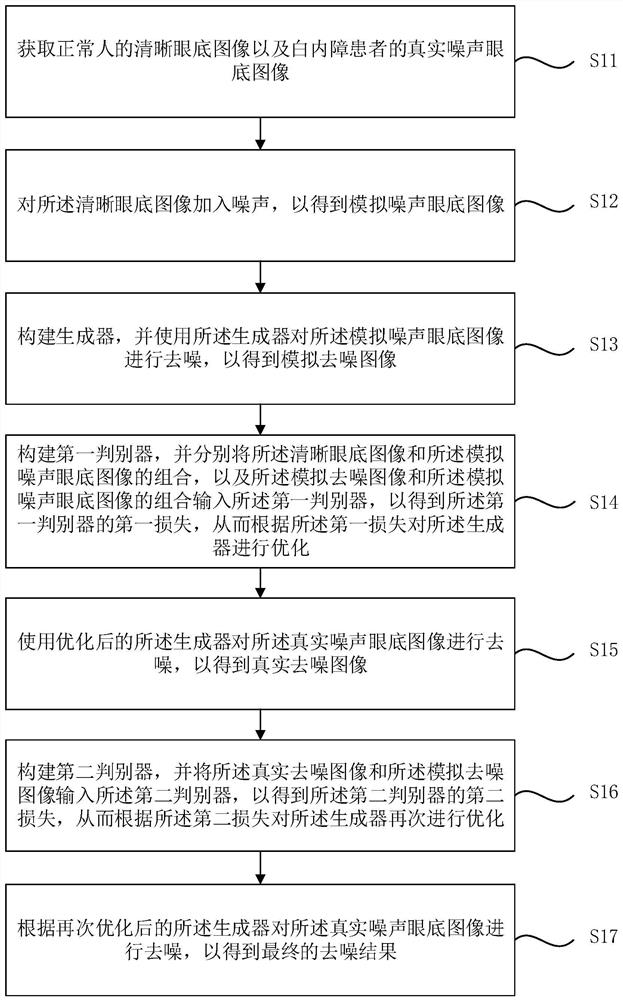
[0033] figure 1 It is a flowchart of a method for denoising a fundus color photo image of a cataract patient provided by Embodiment 1 of the present invention. This embodiment is applicable to the situation where denoising is performed only based on the real noisy fundus images of cataract patients before surgery to better formulate a surgical plan. Execution, the device can be implemented by hardware and / or software, and generally can be integrated into computer equipment. Such as figure 1 As shown, it specifically includes the following steps:

[0034] S11. Obtain a clear fundus image of a normal person and a real noisy fundus image of a cataract patient.

[0035] Specifically, the method provided in this embodiment can make the output of the real noisy fundus image in the denoising model learn the good performance of the simulated noisy fundus image obtained by adding noise to the clear fundus image in the output of the denoising model through domain adaptation , then t...

Embodiment 2

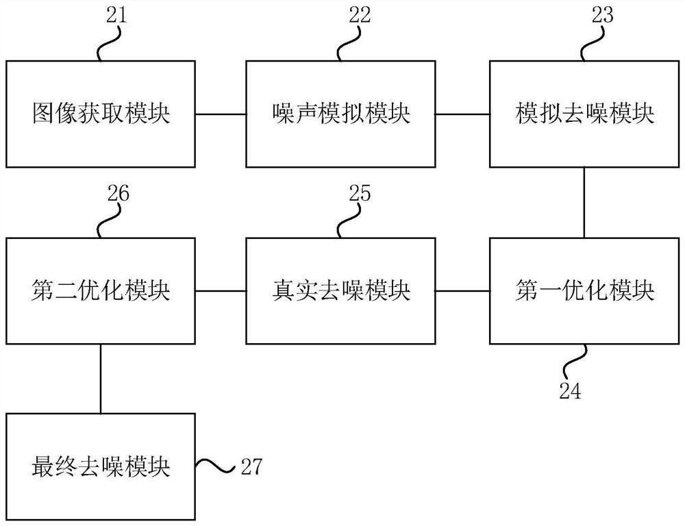
[0064] figure 2 A schematic diagram of the structure of a device for denoising images of fundus photos of cataract patients provided in Embodiment 2 of the present invention. The device can be implemented by hardware and / or software, and can generally be integrated into computer equipment to implement any embodiment of the present invention. Provided is a denoising method for color fundus images of cataract patients. As shown in Figure 2, the device includes:

[0065] The image acquisition module 21 is used to acquire clear fundus images of normal people and real noisy fundus images of cataract patients;

[0066] A noise simulation module 22, configured to add noise to the clear fundus image to obtain a simulated noise fundus image;

[0067] The analog denoising module 23 is configured to construct a generator, and use the generator to denoise the simulated noise fundus image to obtain a simulated denoised image;

[0068] The first optimization module 24 is used to constru...

Embodiment 3

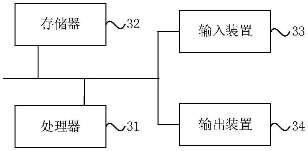
[0092] image 3 The schematic structural diagram of the computer device provided for the third embodiment of the present invention shows a block diagram of an exemplary computer device suitable for implementing the embodiment of the present invention. image 3 The computer equipment shown is only an example, and should not bring any limitation to the functions and scope of use of the embodiments of the present invention. Such as image 3 As shown, the computer equipment includes a processor 31, a memory 32, an input device 33 and an output device 34; the number of processors 31 in the computer equipment can be one or more, image 3 Taking a processor 31 as an example, the processor 31, memory 32, input device 33 and output device 34 in the computer equipment can be connected by bus or other methods, image 3 Take connection via bus as an example.

[0093] The memory 32, as a computer-readable storage medium, can be used to store software programs, computer-executable progra...


PUM


Abstract

The embodiment of the invention discloses a method and a device for denoising a fundus color photo image of a cataract patient, equipment and a medium. The method comprises the following steps: acquiring a clear eye fundus image of a normal person and a real noise eye fundus image of the cataract patient; adding noise to the clear fundus image to obtain a simulated noise fundus image; constructing a generator and denoising the simulated noise eye fundus image by using the generator to obtain a simulated denoised image; constructing a first discriminator, respectively inputting the combination of the simulated noise fundus image, the clear fundus image and the simulated de-noised image to obtain the loss of the first discriminator, and optimizing the generator according to the loss; denoising the real noise eye fundus image by using the optimized generator to obtain a real denoised image; constructing a second discriminator, inputting the real denoised image and the simulated denoised image to obtain the loss of the second discriminator, and optimizing the generator according to the loss; and denoising the real noise eye fundus image according to the re-optimized generator to obtain a final denoising result. Effective denoising of the eye fundus image of the cataract patient is realized.

Description

technical field [0001] The embodiments of the present invention relate to the technical field of medical detection, and in particular to a method, device, equipment and medium for denoising a color fundus photo image of a cataract patient. Background technique [0002] Cataracts, caused by decreased transparency of the lens of the lens, are the leading cause of blindness worldwide. Since there is no effective drug in the clinical treatment of cataract at present, surgery is the only effective treatment. However, cataract surgery will cause damage to the eyes. In the case of other fundus diseases, the risk of surgery and postoperative recovery will be affected. For clinical cases of cataract with fundus lesions, it is necessary to formulate a targeted surgical plan according to the patient's condition to assess the surgical risk and predict the surgical effect. However, due to the opacity of the lens of cataract patients, there will be noise interference during the image sh...

Claims


Application Information

Patent Timeline
IPC IPC(8): G06T5/00G06T5/50G06N3/04G06N3/08
CPCG06T5/50G06N3/08G06T2207/10024G06T2207/20081G06T2207/20084G06T2207/30041G06N3/045G06T5/70
Inventor 李衡刘浩锋刘江胡衍
Owner SOUTH UNIVERSITY OF SCIENCE AND TECHNOLOGY OF CHINA
Learn More