Detection method of content of neutralizing antibody of adeno-associated virus and construction method of cell line

A construction method and detection method technology, applied in the field of medical detection, can solve the problems of inability to meet the real-time monitoring of stable cell lines, long construction period, false positives of cell lines, etc., so as to avoid the problem of drug-resistant bacteria and shorten the construction time.

Pending Publication Date: 2021-09-24
SICHUAN UNIV
10 Cites 0 Cited by

AI-Extracted Technical Summary

Problems solved by technology

[0002] At present, the construction method of adeno-associated virus-infected sensitive cell lines has problems such as long construction...

Method used

AAVR (AAV receptor, AAVR) is a receptor that plays a key role in the AAV infection process, and the infection of a wide range of AAV serotypes is promoted, because AAVR is a general receptor, so It can be applied to the detection of AAV neutralizing antibodies of various serotypes. By constructing a cell line overexpressing the miniAAVR-mCherry protein, the detection threshold, sensitivity and reliability can be improved.
[0037] Since the amount of AAVR protein expressed by animal cells is not high enough, the cells are not sensitive enough to ...

Abstract

The invention discloses a detection method of the content of a neutralizing antibody of an adeno-associated virus and a construction method of a cell line, and relates to the technical field of medical detection. The detection method comprises the following steps of: infecting cells with an overexpression vector of a miniAAVR-fluorescence reporter gene fusion gene, and overexpressing a miniAAVR-fluorescence reporter gene to obtain a cell line sensitive to adeno-associated virus infection. By utilizing the construction method provided by the invention, various animal cells can be quickly transformed into the cell line sensitive to the adeno-associated virus. As the method does not need repeated screening of antibiotics, the construction time of the cell line can be greatly shortened. Meanwhile, the fluorescence reporter gene is used based on the screening process, so that no antibiotic marker is used, false positive stable cell strains caused by drug resistance of the cells can be avoided, and the required stable cell line can be quickly screened.

Application Domain

Technology Topic

Adeno-associated virusAntibody +10

Image

  • Detection method of content of neutralizing antibody of adeno-associated virus and construction method of cell line
  • Detection method of content of neutralizing antibody of adeno-associated virus and construction method of cell line
  • Detection method of content of neutralizing antibody of adeno-associated virus and construction method of cell line

Examples

  • Experimental program(6)
  • Comparison scheme(1)

Example Embodiment

[0059] Example 1
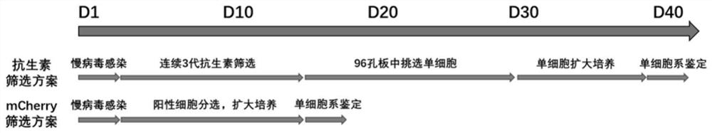
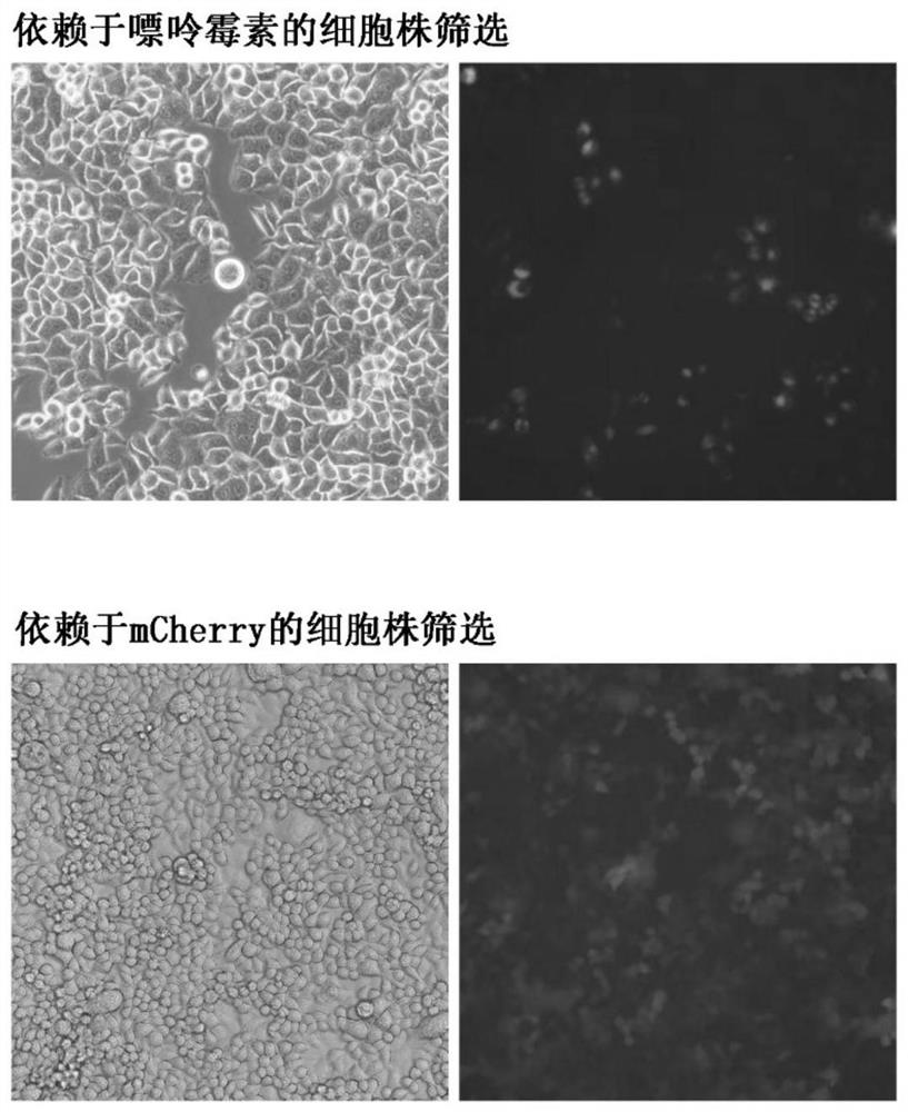
[0060] This embodiment provides a method for constructing a cell line sensitive to adeno-associated virus infection, and the specific construction steps are as follows:
[0061] Introduce the lentiviral vector overexpressing the miniAAVR-mCherry gene into Hela cells; obtain Hela cells overexpressing miniAAVR-mCherry, that is, Hela cells sensitive to adeno-associated virus infection;
[0062] Adeno-associated virus-infected sensitive Hela cells were cultured to obtain a large number of adeno-associated virus-infected sensitive Hela cells.
[0063] Specifically, the construction process of the lentiviral vector of miniAAVR-mCherry gene is as follows (linear sequence refers to image 3 shown):
[0064] (1) Original plasmid sequence:
[0065] AAVR cDNA: Extract RNA from hela cells, reverse transcribe cDNA, use this as a template, and PCR produce AAVRcDNA.
[0066] Vector sequence: the vector is pLVX-Puro vector, purchased from transvector;
[0067] mCherry sequence: CDS region from laboratory plasmid pAAV-CB-mCherry
[0068] (II) Construction process:
[0069] (1) Restriction site selection: select BamH I and Xba I on the vector as the inserted restriction site;
[0070] (2) Primer design: Use overlap extension PCR to amplify three small fragments respectively (for the amplification primers, refer to the F-terminal primers and R-terminal primers of the F1 fragment, F2 fragment and F3 fragment shown in the table below), and then PCR tandem into a complete insert fragment (namely miniAAVR-mCherry gene).
[0071]
[0072]
[0073] (3) Preparation of PCR system: Q5 ultra-fidelity polymerase is used, and GCenhancer can be added when the gene GC content is high. The PCR reaction system is as follows:
[0074] Reagent name 25ul system sample volume Final concentration 5xQ5 Reaction Buffer 5ul 1x dNTP (10mM) 0.5ul 200uM Primer-F(10uM) 1.25ul 0.5Um Primer-R(10uM) 1.25ul 0.5Um template DNA variable less than 1000ng Q5 hot start enzyme 0.25ul 0.02U/ul 5Xq5 GC enhancer 5ul (optional) 1x wxya 2 o
[0075] The PCR reaction procedure is as follows:
[0076]
[0077] (4) Acquisition of the vector and the inserted gene: the overexpression vector is the lentiviral vector PLVX-Puro, which is transformed into Escherichia coli DH5α for amplification. The target gene was obtained by the above PCR steps.
[0078] (5) Carrier and target gene digestion: use NEB restriction endonucleases to digest the target gene and vector according to the designed restriction site. The restriction system is as follows:
[0079] Reagent name Adding amount 10x buffer 5ul DNA (plasmid or PCR fragment) more than 2ug restriction endonuclease 0.5ul/each enzyme ddH 2 O
[0080] The 10xbuffer and reaction temperature vary depending on the enzyme. For details, see the recommended reaction conditions on the official website.
[0081] (6) Enzymatic fragment recovery: Add 6x loading buffer to the digested system to stop, run the gel at 150V for 30 minutes for recovery, and use the gel recovery kit to recover the target DNA fragment after cutting the gel. The glue recovery steps are as follows:
[0082] (a) Weigh, add the same volume of DNA binding buffer, melt at 55°C for 10 minutes, invert and mix;
[0083] (b) liquid gel recovery column, centrifuged at 10000g for 1 minute;
[0084] (c) Discard the liquid in the recovery tube, add 300ul DNA binding buffer to the recovery column, and centrifuge at 10000g for 1 minute;
[0085] (d) Discard the liquid, add 700ul DNA wash buffer, and centrifuge at 10000g for 1 minute;
[0086] (e) repeat step d;
[0087] (f) Centrifuge at 13000g for 2 minutes to completely remove alcohol residue, and transfer the column to a new EP tube;
[0088] (g) Add 30ul Elution buffer to the recovery column, let stand for 1 minute, and then centrifuge at 13000g for 1 minute.
[0089] (7) Vector ligation: The vector and the insert were ligated according to the following system.
[0090] Reagent name Adding amount 10xT4 ligase buffer 1ul T4 DNA ligase 0.5ul vector fragment 1ul insert Vector: Insert = 1:3 (molar ratio) ddH 2 O
[0091] The reaction temperature is 16°C, and the connection time is not less than 1 hour.
[0092](8) Ligation product conversion. Melt 50ul Escherichia coli competent (DH5α) on ice, add 5ul ligation product, place on ice for 30 minutes, then heat shock at 42°C for 30-45s, after two minutes in ice bath, add 300ul non-antibiotic medium, 220rpm, 37 Incubate on a shaker at °C for 1 hour.
[0093] (9) Plate coating: after culture, take 300 ul of the culture solution, evenly spread it on the plate containing the corresponding antibiotics, turn it upside down, and incubate overnight at a constant temperature of 37°C.
[0094] (10) Selection of positive clones
[0095] About 16 hours after plating, pick 2-4 cloned colonies and put them into the liquid medium with corresponding antibiotics, culture at 220 rpm, 36°C for 16-18 hours with shaking.
[0096] (11) Plasmid extraction
[0097] According to the instructions of the omega plasmid mini-extraction kit, carry out the plasmid extraction, the specific steps are as follows:
[0098] (a) Collect 1ml of the bacterial solution into an EP tube, centrifuge at 10000g for 1min, and remove the supernatant;
[0099] (b) Add 250ul of solutionI with RNase to resuspend the bacteria;
[0100] (c) Add 250ul solutionII and mix well;
[0101] (d) Add 350ul solutionIII, mix immediately, and centrifuge at 13000g for 10min;
[0102] (e) After putting the extraction column into the corresponding collection tube, add the supernatant from the previous step to the column, centrifuge at 10000g for 1 minute, and discard the liquid in the collection tube;
[0103] (f) Add 500ul of HEB buffer with isopropanol to the column, centrifuge at 10000g for 1 minute, discard the liquid in the collection tube;
[0104] (g) Add 700ul of Wash buffer with absolute ethanol to the column, centrifuge at 10000g for 1 minute, and discard the liquid in the collection tube;
[0105] (h) repeat step 7;
[0106] (i) The column is put into a collection tube, and idling at 13000g for 2 minutes to remove residual ethanol;
[0107] (j) Discard the collection tube, put the column into a new EP tube, add 50ul Elution buffer to elute the DNA, let it stand at room temperature for 1 minute, and then centrifuge at 13000g for 1 minute to obtain a plasmid solution with a volume of about 50ul.
[0108] (12) Identification of positive clones:
[0109] Select the corresponding enzyme according to the gene and vector sequence for positive plasmid identification, and prepare the following 10ul plasmid identification system:
[0110]
[0111]
[0112] According to the selected reaction buffer and optimal temperature of the enzyme, the reaction time of enzyme digestion identification is more than 30 minutes, and then the gel is run to determine whether the enzyme digestion result is correct.
[0113] Of course, in other embodiments, the AAVR gene, AAVR homologous gene, or AAVR gene fragments of Hela cells can be knocked out or disturbed first, and then the expression vector that overexpresses miniAAVR-mCherry can be introduced; in addition, pancreatic islets can also be used Cells, CHO cells, 293 cells, HepG2 cells and other types of cells were tested, and then the lentiviral vector overexpressing the miniAAVR-mCherry gene was introduced to obtain the corresponding adeno-associated virus-sensitive cell lines.

Example Embodiment

[0114] Example 2
[0115] This embodiment provides a method for detection and content determination of adeno-associated virus neutralizing antibodies, and the specific operation steps are as follows:
[0116] The adeno-associated virus infection-sensitive Hela cells constructed in Example 1 were cultured in a 24-well plate, and the number of cells was 1×10 5 Cells/well density were plated;
[0117] Dilute the sample to be tested in a 10-fold concentration gradient, the highest dilution is 1×10 9 times, to obtain 10 different concentrations of samples to be tested;
[0118] And set the maximum value control group (do not contain AAV antibody, add neonatal umbilical cord blood serum to replace the antibody sample) and minimum value control group (do not add AAV during detection, use DMEM to replace AAV);
[0119] Take 10 samples of different concentrations to be tested, the maximum value control group and the minimum value control group, a total of 12 groups, take 10 μL respectively, mix and incubate with 10 μL recombinant adeno-associated virus solution with GFP gene, and add to each sample The amount of recombinant adeno-associated virus with GFP gene is 10 20 Virus particles/ml; Incubate for 75 minutes at an incubation temperature of 45°C to obtain a mixed sample;
[0120] The mixed samples were infected with Hela cells sensitive to adeno-associated virus infection and cultured at 37°C.
[0121] After 48 hours of infection, the GFP fluorescence signal of the infected cells was detected by flow cytometry, and compared with the signal values ​​of the maximum value control group and the minimum value control group, respectively.
[0122] Of course, in other embodiments, genes such as EGFP gene, YFP gene, LacZ gene, GUS gene, and luciferase gene can also be selected as characteristic genes for detection.

Example Embodiment

[0123] Example 3
[0124] This embodiment provides a method for detection and content determination of adeno-associated virus neutralizing antibodies, and the specific operation steps are as follows:
[0125] (1) The adeno-associated virus infection-sensitive Hela cells constructed in Example 1 were cultured in a 24-well plate, and the number of cells was 1×10 5 Cells/well density were plated;
[0126] (2) Dilute the sample to be tested in a 10-fold concentration gradient, the highest dilution is 1×10 9 times, to obtain 10 different concentrations of samples to be tested;
[0127] (3) And set the maximum value control group (without AAV antibody, adding neonatal umbilical cord blood serum to replace the antibody sample) and the minimum value control group (do not add AAV during detection, use DMEM instead of AAV);
[0128] (4) Take 10 different concentrations of samples to be tested, the maximum value control group and the minimum value control group, a total of 12 groups, take 30 μL respectively, and incubate with 10 μL of recombinant adeno-associated virus solution with GFP gene respectively. The amount of recombinant adeno-associated virus with GFP gene added to the sample is 1 virus particle/ml; incubate for 30 minutes at a shaker temperature of 60°C to obtain a mixed sample;
[0129] (5) Infect the mixed sample with the sensitive Hela cells infected with the adeno-associated virus in step 1.1, and culture at 37°C.
[0130] (6) After 48 hours of infection, the GFP fluorescence signal of the infected cells was detected by flow cytometry, and compared with the signal values ​​of the maximum value control group and the minimum value control group, respectively.
[0131] Of course, in other embodiments, genes such as EGFP gene, YFP gene, LacZ gene, GUS gene, and luciferase gene can also be selected as characteristic genes for detection.

PUM

no PUM

Description & Claims & Application Information

We can also present the details of the Description, Claims and Application information to help users get a comprehensive understanding of the technical details of the patent, such as background art, summary of invention, brief description of drawings, description of embodiments, and other original content. On the other hand, users can also determine the specific scope of protection of the technology through the list of claims; as well as understand the changes in the life cycle of the technology with the presentation of the patent timeline. Login to view more.

Similar technology patents

Rapid construction method of program white list

ActiveCN114579967AReduced build timeDigital data authenticationPlatform integrity maintainanceWhitelistInstruction step
Owner:北京珞安科技有限责任公司

Epidermis skin model with melanin and construction method and application of epidermis skin model

PendingCN111117945ASimplify the operation processReduced build timeCulture processEpidermal cells/skin cellsStratum basaleCell layer
Owner:GUANGDONG BOXI BIO TECH CO LTD

Establishment method of three-dimensional cell model

PendingCN105671029AEasy to operateReduced build timeCulture processElectrical/wave energy microorganism treatmentBiologyFerromagnetism
Owner:THE SIXTH AFFILIATED HOSPITAL OF SUN YAT SEN UNIV

Gingival epithelium model and in-vitro building method thereof

InactiveCN107312743AGood repeatabilityReduced build timeCulture processEpidermal cells/skin cellsPhases of clinical researchStructure and function
Owner:GUANGDONG BOXI BIO TECH CO LTD

Classification and recommendation of technical efficacy words

Why Eureka
Try Eureka