
Skin lesion segmentation method based on convolution attention model

An attention model, a technology for skin lesions, applied in neural learning methods, biological neural network models, image analysis, etc., can solve the problems of incomplete lesion area, large amount of parameters, imperfect feature extraction, etc., to improve feature representation, The effect of improving performance

Pending Publication Date: 2021-12-03
NORTHWEST NORMAL UNIVERSITY
View PDF0 Cites 5 Cited by
  • Summary
  • Abstract
  • Description
  • Claims
  • Application Information

AI Technical Summary

Problems solved by technology

However, in the field of skin lesion segmentation, due to the blurred boundaries of the skin lesion area, the lesions of different subjects show obvious differences in location, shape, and color, as well as a large number of artifacts, including inherent skin features (such as hair, blood vessels) and artificial The existence of artifacts (such as air bubbles, ruler marks, uneven illumination, incomplete lesion areas, etc.) makes it still a great challenge for general convolutional neural networks to accurately segment the boundaries of skin lesions
The previous literature has solved the above problems to a certain extent. However, the skin surface images used in the model come from patient skin surface photos rather than publicly available public data sets, which cannot better verify the performance of the model.
Secondly, the convolutional neural network structure, as the depth increases, the data information will continue to be lost, and the imperfect feature extraction will lead to poor model segmentation performance
Repeated convolution operations can enhance the feature extraction, but this process is likely to cause too many parameters, and data calculation redundancy further causes the model to overfit, which greatly reduces the prediction accuracy of the model.

Method used


Image

  • Skin lesion segmentation method based on convolution attention model
  • Skin lesion segmentation method based on convolution attention model
  • Skin lesion segmentation method based on convolution attention model

Examples


Embodiment 1

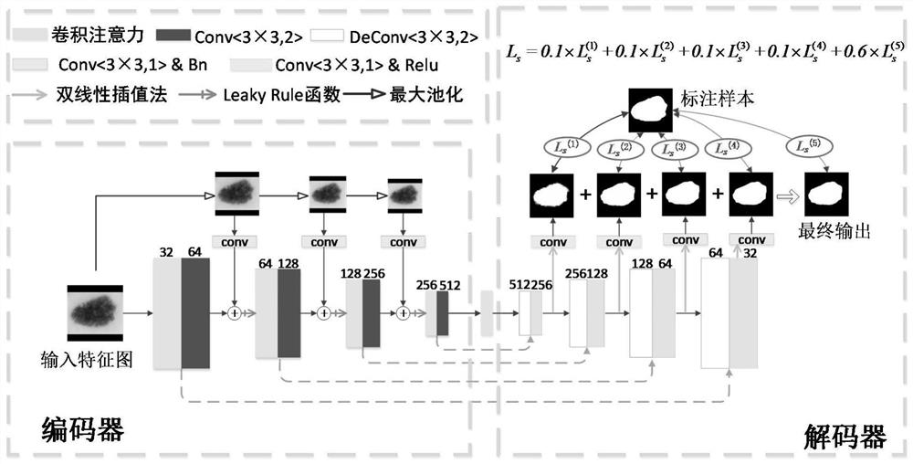
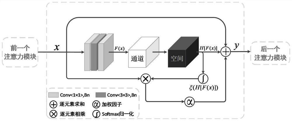
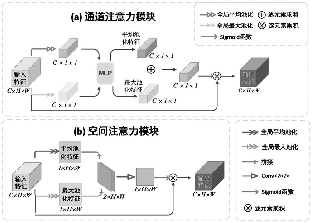
[0043] Such as Figure 1-5 As shown, the present invention provides a kind of skin lesion segmentation method based on convolutional attention model, and this segmentation method specifically follows the steps:

[0044] Step 1: Select ISIC-2017 dataset and PH2 dataset:

[0045] The ISIC-2017 dataset contains 8-bit RGB dermoscopy images ranging in size from 540×722-4499×6748 pixels. The dataset provides 2000 training images, a separate dataset of 150 images for validation, and a separate dataset for A separate dataset of 600 images was tested. All dermoscopy images in the dataset were labeled as benign moles, melanomas, or seborrheic keratoses. The PH2 dataset contained 200 images, of which 160 were moles. are common moles and atypical moles, and the remaining 40 images are melanomas. The images in the PH2 dataset are 8-bit RGB images with a fixed size of 768×560 pixels, collected under the same conditions with a magnification of 20 times;

[0046] Step 2: Data preprocessing:...


PUM


Abstract

The invention discloses a skin lesion segmentation method based on a convolution attention model, and the method specifically comprises the following steps: 1, selecting an ISIC-2017 data set and a PH2 data set which comprise 8-bit RGB dermatoscopy images, wherein the image size is from 540 * 722 to 4499 * 6748 pixels, the data set provides 2000 training images, an independent data set of 150 images used for verification and an independent data set of 600 images used for testing, all dermatoscopy images in the data set are marked as benign nevus, melanoma or seborrheic keratosis respectively, and the PH2 data set comprises 200 images, wherein 160 images are nevus and are divided into common nevus and atypical nevus. The method is used for accurate skin lesion segmentation in dermatoscopy images, and has practical reference value for medical researchers. A multi-scale input module is added to the model, image features are extracted by using a convolution attention module, parameters are updated through a multi-label loss function to train the model, and a final segmentation image is generated.

Description

technical field [0001] The invention relates to the field of computer vision medical image processing, in particular to a skin lesion segmentation method based on a convolutional attention model. Background technique [0002] As we all know, skin, as the largest organ of the human body, is usually directly exposed to the air, making skin diseases one of the most common diseases in humans. Melanoma is the most lethal malignant skin tumor, causing more than 10,000 deaths per year. However, if caught early, melanoma can be cured with simple excision. However, in actual diagnosis, melanoma cannot be detected correctly based on perception and vision alone, even when dermoscopy is performed by experienced dermatologists. Computer-aided analysis avoids many of these problems and is increasingly being studied to help dermatologists improve the efficiency and objectivity of dermoscopic image analysis. Automatic segmentation of skin lesions is an important step in computer-aided de...

Claims


Application Information

Patent Timeline
IPC IPC(8): G06T7/00G06T7/11G06T7/90G06T3/40G06K9/62G06N3/04G06N3/08
CPCG06T7/0012G06T7/11G06T7/97G06T3/4038G06T7/90G06N3/084G06T2207/30088G06N3/045G06F18/214
Inventor 蒋芸曹思敏陶生鑫吴超刘文欢
Owner NORTHWEST NORMAL UNIVERSITY
Learn More