Monitoring method of antibody coupled latex microspheres and application thereof

A technology of antibody coupling and latex, which is applied in the field of immunoassay medical detection, can solve the problems of affecting detection results and low coupling efficiency

Pending Publication Date: 2022-04-01
NANJING VAZYME MEDICAL TECH CO LTD
View PDF6 Cites 0 Cited by
  • Summary
  • Abstract
  • Description
  • Claims
  • Application Information

AI Technical Summary

Problems solved by technology

[0004] If the coupling efficiency is not high, when the antigen to be detected in the system is insufficient, there will be many reactive groups on the surface of the latex microspheres after coupling, and these groups can react with antibodies that have been coupled to other microspheres. As a result, a non-detectable agglutination reaction occurs, which affects the final detection result

Method used


Image

  • Monitoring method of antibody coupled latex microspheres and application thereof
  • Monitoring method of antibody coupled latex microspheres and application thereof
  • Monitoring method of antibody coupled latex microspheres and application thereof

Examples


Embodiment 1

[0051] Example 1: Preparation of antibody-latex microsphere coupling complex

[0052] (1) Prepare 0.02-0.05mol / L MES buffer solution, adjust the pH to 6.00-6.50 with 0.05mol / L sodium hydroxide solution, and mix thoroughly;

[0053] (2) Weigh 0.2383g of HEPES solid and dissolve it in 100ml of 0.1M, pH 3.5 sodium citrate aqueous solution;

[0054] (3) Take a certain amount of adiponectin (NADP) monoclonal antibody NA1, dilute to 2 mg / ml with the buffer in (2), and set aside;

[0055] (4) Take 2ml of latex microspheres with a particle size of 123nm and a solid content of 5%, dilute to 100ml with the solution in (1), stir and mix, and shake at a constant temperature of 37°C for 30 minutes;

[0056] (5) The activator needs to be prepared and used immediately. Weigh 0.2490g of EDC solid, make it into 1mg / ml with purified water, and then add it to the reaction solution of (4), place on a shaker, and shake at a constant temperature of 37°C for 30 minutes;

[0057] (6) Take 0.5ml of ...

Embodiment 2

[0059] Example 2: Monitoring of antibody-latex microsphere coupling process

[0060] (1) After adding the diluted antibody solution obtained in (3) in step (7) of Example 1, sample 200ul at different time points of the reaction, and the sampling time points are 10min, 20min, 30min, 40min, 50min, 60min. Under the conditions of 16000rpm and 15min, centrifuge each sample with a centrifuge, remove the supernatant and resuspend the centrifuged product with purified water, and detect the fluorescence signal value at each sampling time point with a UV spectrophotometer, and the UV detection wavelength is 565nm.

Embodiment 3

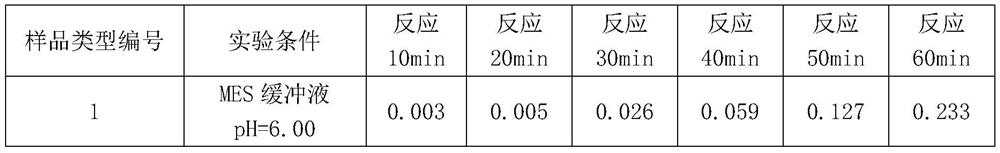
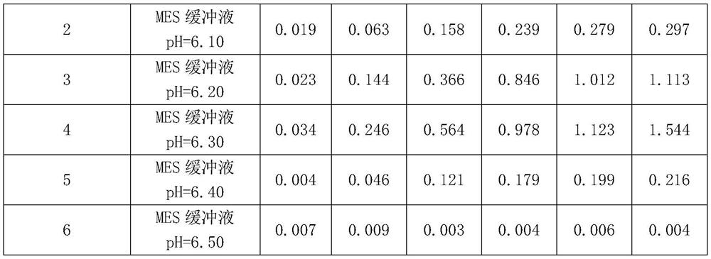
[0061] Example 3: Monitoring of antibody-latex microsphere coupling process under different pH MES buffer conditions

[0062] Select 0.05mol / L MES buffer solution with different pH (pH is 6.00, 6.10, 6.20, 6.30, 6.40, 6.50 respectively), and prepare the antibody-latex microsphere coupling complex according to the method of Example 1, according to Example 2 The method was used to measure the fluorescence signal value of the resuspension solution at 10min, 20min, 30min, 40min, 50min, and 60min, so as to evaluate the coupling efficiency.

[0063] The experimental results (Table 1) show that changing the pH of the MES buffer will directly affect the coupling efficiency; but no matter how much the pH of the MES buffer is, within 60 minutes after the start of the reaction, the fluorescence signal value increases with the reaction time increases, indicating that the coupling efficiency is proportional to the reaction time.

[0064] According to Table 1, when the reaction is 10min an...


PUM

PropertyMeasurementUnit
particle diameteraaaaaaaaaa
diameteraaaaaaaaaa
particle diameteraaaaaaaaaa

Abstract

The invention discloses a coupling process monitoring method for antibody coupling latex microspheres, and belongs to the field of immunoassay medical detection. According to the invention, antibody labeling is carried out by utilizing the adsorbability of the fluorescent dye to the antibody, then the uncoupled antibody in the solution is removed, and the real-time monitoring on the coupling efficiency change in the coupling process is realized by observing the change of the fluorescence signal value of the latex microsphere-fluorescence labeled antibody compound at different reaction times; the reaction condition with the optimal coupling efficiency can be found.

Description

technical field [0001] The invention belongs to the field of immunoanalysis medical detection. Specifically, the present invention relates to a monitoring method and application of antibody-coupled latex microspheres. Background technique [0002] Latex immunoturbidimetry is a stable and accurate method for the homogeneous immunoturbidimetric detection of body fluid proteins. It has two detection methods: scattering turbidimetry and transmission turbidimetry. The basic principle of these two methods is that latex microspheres physically adsorb antibodies. When the adsorbed antibodies bind to antigens, they quickly aggregate in a short time, thereby changing the astigmatism or light transmission properties of the reaction solution. [0003] The latex-enhanced immunoturbidimetric assay (particle-enhanced turbidimetric immunoassay, PETIA) uses chemical coupling to connect microspheres and antibodies to form complexes, which then bind to antigens and aggregate. Compared with p...

Claims


Application Information

Patent Timeline
Patent Type & Authority Applications(China)
IPC IPC(8): G01N33/533G01N33/543G01N33/577G01N33/74G01N21/64
Inventor 唐波时亚斌赖迪文吴凡
Owner NANJING VAZYME MEDICAL TECH CO LTD
Why Eureka
Try Eureka