
Nasopharyngeal carcinoma positioning segmentation method and system based on image segmentation convolutional neural network

A convolutional neural network and image segmentation technology, applied in the field of medical diagnosis, can solve the problem of inability to judge malignant tumors, and achieve the effect of improving the treatment effect, improving the accuracy and improving the detection rate.

Pending Publication Date: 2022-04-19
THE FIRST AFFILIATED HOSPITAL OF SUN YAT SEN UNIV +1
View PDF1 Cites 5 Cited by
  • Summary
  • Abstract
  • Description
  • Claims
  • Application Information

AI Technical Summary

Problems solved by technology

[0005] However, the current artificial intelligence nasal endoscopy-assisted diagnosis system is a binary classification diagnosis model. The system judges the nasopharynx image as "malignant tumor" or "non-malignant tumor" by calculating the probability of nasal endoscopic images. As a result, it is impossible to make an exact judgment on the exact location of the malignant tumor in the image

Method used


Image

  • Nasopharyngeal carcinoma positioning segmentation method and system based on image segmentation convolutional neural network
  • Nasopharyngeal carcinoma positioning segmentation method and system based on image segmentation convolutional neural network
  • Nasopharyngeal carcinoma positioning segmentation method and system based on image segmentation convolutional neural network

Examples


Embodiment Construction

[0020] The specific embodiments of the present invention will be further described below in conjunction with the accompanying drawings. It should be noted here that the descriptions of these embodiments are used to help understand the present invention, but are not intended to limit the present invention. In addition, the technical features involved in the various embodiments of the present invention described below may be combined with each other as long as they do not constitute a conflict with each other.

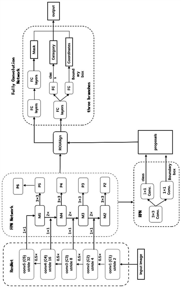
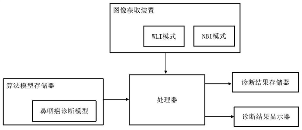
[0021] The nasopharyngeal carcinoma positioning and segmentation method and system based on image segmentation convolutional neural network, combined with the existing electronic nasal endoscopy system equipped with WLI and NBI modes, can analyze the nasopharyngeal endoscopic images taken by the operator in real time, And output the electronic nasal endoscopic image segmented by the auxiliary diagnosis system (that is, stroke the malignant lesion area), prompting the nat...


PUM


Abstract

The invention discloses a nasopharyngeal carcinoma positioning segmentation method and system based on an image segmentation convolutional neural network, and the method comprises the steps: obtaining an electronic nose endoscopic image through employing a WLI mode and an NBI mode, inputting the electronic nose endoscopic image into a nasopharyngeal carcinoma diagnosis model based on the image segmentation convolutional neural network, obtaining a malignant tumor region marked by the diagnosis model, and carrying out the positioning segmentation of the nasopharyngeal carcinoma. The diagnosis system can judge the captured image in real time, mark the malignant tumor part in the nasopharyngeal carcinoma image, export the diagnosis result, intuitively judge whether the target lesion is the malignant tumor tissue or not and determine the boundary range of the malignant tumor lesion according to the malignant tumor lesion as long as the lens is focused on the suspicious lesion tissue in the nasopharyngeal cavity. Suspicious diseased regions are quickly selected for biopsy, so that the accuracy of nasopharyngeal carcinoma detection under a nasal endoscope is effectively improved, and the detection rate of biopsy is increased.

Description

technical field [0001] The invention belongs to the field of medical diagnosis, and in particular relates to a method and system for diagnosing the nature and scope of nasopharyngeal carcinoma by using artificial intelligence to identify a nasal endoscopic image and capture a malignant tumor target region (ROI). Background technique [0002] Nasopharyngeal carcinoma (NPC) refers to malignant tumors that occur on the top and side walls of the nasopharyngeal cavity, and is one of the high-risk malignant tumors. The current electronic nasal endoscopy uses ordinary white light imaging (WLI) as the lighting device, which has certain limitations in clarity and contrast, and it is easy to miss the diagnosis of superficial early cancer and precancerous lesions that occur on the mucosal surface. Narrow band imaging (narrow band imaging, NBI) technology uses a narrow-spectrum filter to remove the red light in the ordinary endoscope, and only releases two wavelengths of light with a ce...

Claims


Application Information

Patent Timeline
Patent Type & Authority Applications(China)
IPC IPC(8): G06T7/00G06T7/11G06T7/73G06V10/764G06V10/82G06K9/62G06N3/04G06N3/08
CPCG06T7/0012G06T7/11G06T7/73G06N3/08G06T2207/10068G06T2207/30096G06N3/048G06N3/045G06F18/24
Inventor 文译辉龙宇栋项毅帆雷文斌文卫平林浩添肖钧
Owner THE FIRST AFFILIATED HOSPITAL OF SUN YAT SEN UNIV
Learn More