System and method for determining three-dimensional functional liver segment based on medical image

A medical imaging and imaging technology, applied in the field of computer vision medical image processing, can solve the problems of not being able to provide accurate location information of liver function distribution, not being able to provide information on the spatial distribution of liver function, and not being able to directly apply radiation therapy as a result, so as to reduce the total treatment cost. The effect of increasing the number of times, ensuring the integrity of liver function, and reducing the cost of treatment

Pending Publication Date: 2022-08-09
SHANDONG TUMOR HOSPITAL
View PDF0 Cites 3 Cited by
  • Summary
  • Abstract
  • Description
  • Claims
  • Application Information

AI Technical Summary

Problems solved by technology

Liver function blood chemistry tests can effectively evaluate liver metabolism, immunity, synthesis and other functions, but there are limitations in that they cannot provide information on the spatial distribution of liver function
Although liver function imaging can clearly display the distribution of liver function in the liver and realize quantitative assessment of liver function, it still has the problem that it cannot provide anatomical information and precise location information of liver function distribution. Low resolution, difficult to fuse with other anatomical images
Although the existing liver segment segmentation methods can obtain the liver segment segmentation results, the results cannot be directly applied to radiotherapy, and the segmentation results are obtained only by the training parameters of the segmentation network results, without further automatic correction and consideration of the liver segment itself in the population. It is difficult to apply to all liver images and the scope of application is narrow

Method used


Image

  • System and method for determining three-dimensional functional liver segment based on medical image
  • System and method for determining three-dimensional functional liver segment based on medical image
  • System and method for determining three-dimensional functional liver segment based on medical image

Examples


Embodiment Construction

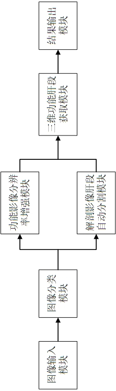
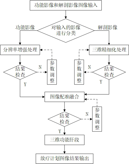
[0060] example, as figure 1 As shown, the system for determining three-dimensional functional liver segments based on medical images includes an image input module, an image classification module, a functional image resolution enhancement module, an anatomical image liver segment automatic segmentation module, a three-dimensional functional liver segment acquisition module and a result output module.

[0061] The image input module is used to input functional images and anatomical images required for liver segment segmentation. The functional image modalities are SPECT and MRI, and the anatomical image modalities are CT. The format conforms to the DICOM3.0 standard, namely digital imaging and communications in medicine.

[0062] The image classification module is used for determining whether the input is a functional image or an anatomical image according to the header file information of each input image slice.

[0063] The functional impact resolution enhancement module is ...


PUM


Abstract

The system for determining the three-dimensional functional liver segment based on the medical image comprises an image input module, an image classification module, a functional image resolution enhancement module, an anatomical image liver segment automatic segmentation module, a three-dimensional functional liver segment acquisition module and a result output module. The functional influence resolution enhancement module performs resolution enhancement processing on the functional image to obtain a high-resolution enhanced functional image, and the anatomical image liver segment automatic segmentation module performs liver segment automatic segmentation on the anatomical image to obtain a refined three-dimensional liver segment anatomical image. A functional image and an anatomical image are input, the input images are classified, resolution enhancement processing is performed on the functional image, three-dimensional refining processing and registration fusion are performed on the anatomical image to obtain a three-dimensional functional liver segment, and an image result is output and imported into a radiotherapy plan design system for radiotherapy dose calculation. According to the invention, three-dimensional functional liver segment judgment can be realized, the method is used for radiotherapy, the segmentation precision is improved, and the treatment cost of a patient is reduced.

Description

technical field [0001] The invention relates to the field of computer vision medical image processing, in particular to a system and method for determining three-dimensional functional liver segments based on medical images. Background technique [0002] Primary liver cancer (hepatocellular carcinoma, HCC) is a high-incidence malignant tumor in my country with poor prognosis and short survival time. Radiotherapy plays an important role in the treatment of HCC. However, radiotherapy can easily cause radiation-induced liver injury (RILI), and there is a significant correlation between radiotherapy and irradiation dose. important limiting factor for control rates. [0003] Since there is no obvious separation (such as diaphragm) boundary between liver segments, and it is not visible in medical images, it is difficult to accurately divide liver segments. At present, the division of liver segments is mainly based on computed tomography (CT). This is done manually. Specifically,...

Claims


Application Information

Patent Timeline
Patent Type & Authority Applications(China)
IPC IPC(8): G06T7/00G06T17/00G06T7/10G06T7/33G06N3/04G06N3/08
CPCG06T7/0012G06T17/00G06T7/10G06T7/33G06N3/088G06T2207/30056G06T2207/30096G06N3/045
Inventor 仇清涛段敬豪李腾翔尹勇陈进琥王兴莉孙涛吴仕章巩贯忠朱健刘同海佟颖丁佳琳尹菡菁
Owner SHANDONG TUMOR HOSPITAL
Why Eureka
Try Eureka