Full term development of animals from enucleated oocytes reconstituted with adult somatic cell nuclei
A technology of oocytes and somatic cells, used in the preparation of hybrid cells, cells modified by introducing foreign genetic material, fermentation, etc.
- Summary
- Abstract
- Description
- Claims
- Application Information
AI Technical Summary
Problems solved by technology
Method used
Image

Examples
Embodiment 1
[0085] Preparation of Somatic Cells
[0086] In this example, cumulus cells were isolated from mouse oviducts as a source of adult somatic nuclei for injection into enucleated mouse oocytes. The method for obtaining the cloned mice produced in Table 2 and in Series A-D described below is also described in Wakayama, et al. 1998, Nature 394, 369-374.
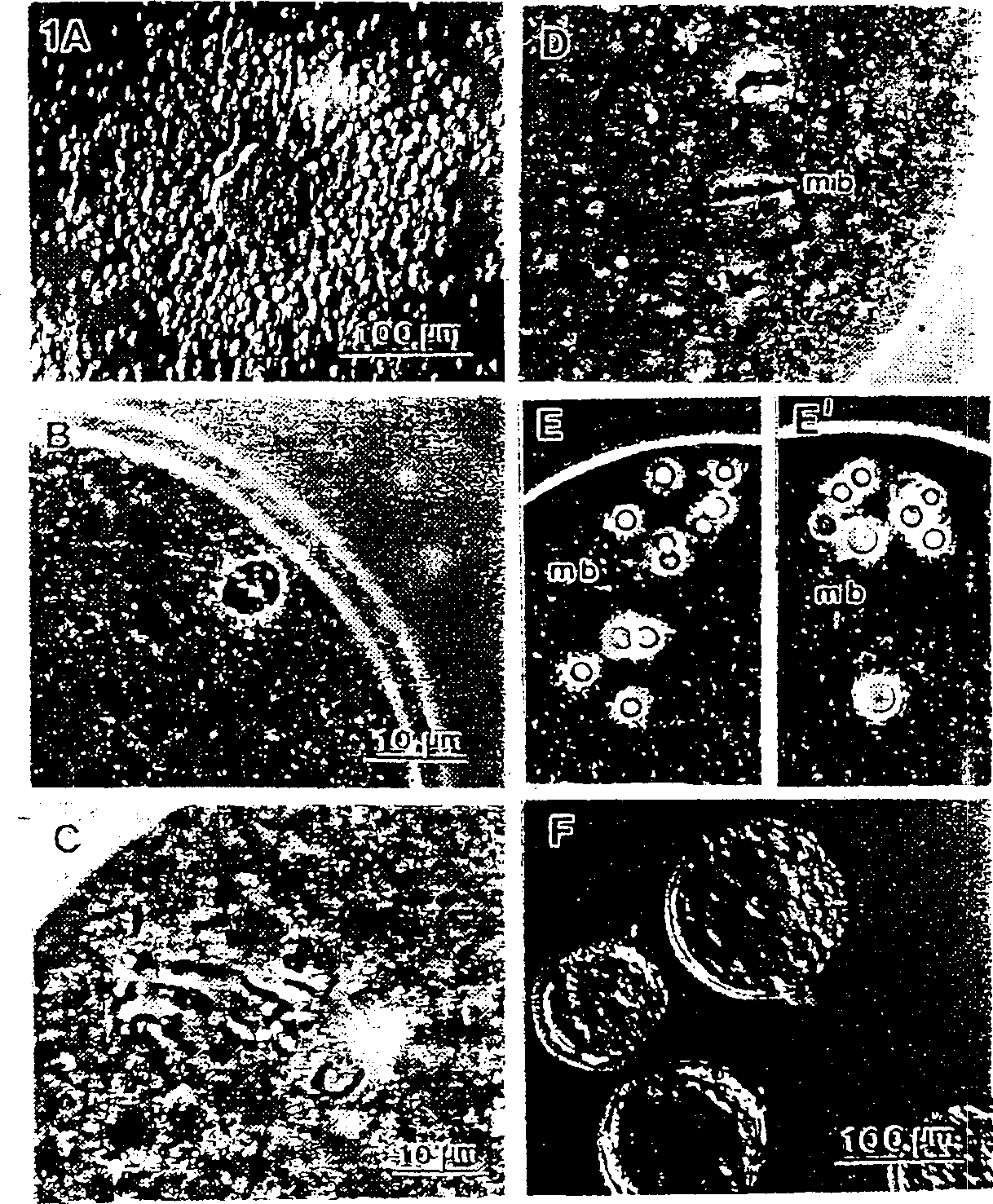
[0087] Female mice B6D2F1 (C57BL / 6×DBA / 2, series A and B), B6C3F1 (C57BL / 6×C3H / He, adopted in series C) or B6C3FI clone mice generated in series D were superovulated. Thirteen hours after hCG injection, cumulus-oocyte complexes were collected from the oviduct (see figure 1 A), cumulus cells were dispersed by treatment in Hepes-CZB medium supplemented with bovine testicular hyaluronidase (0.1% [w / v], 300 units / ml, ICN Biochemicals, Costa Mesa, CA). Medium-sized cumulus cells (10-12 microns in diameter) were the most common (>70%) and were selected for injection. After dispersion, the cells were transferred to Hepes-CZB containi...
Embodiment 2
[0089] Preparation of Somatic Cells
[0090] In this example, podocytes and brain cells (neurons) were isolated from adult mice. These cells are characterized by nondividing in adult animals and are permanently arrested in the G0 phase of the cell cycle.
[0091] Separate the seminiferous tubules from the testes and contact with 1 mg / ml collagenase in Hepes-CZB solution at 30°C for 20 minutes. Vials were then minced with a razor blade and placed in Hepes-CZB containing 1 mg / ml trypsin with occasional agitation. The resulting suspension was then allowed to stand. The podocyte-rich fraction settles first. Aspirate the suspended cells and resuspend the remainder with fresh medium. Podocytes with characteristic morphological features are easily identified under a low power microscope. Manipulation of single podocytes is performed with large injection pipettes (approximately 10 µm inner diameter).
[0092] Neurons were isolated from the cerebral cortex of adult B6D2F1 female ...
Embodiment 3
[0093] The preparation of embodiment 3 somatic cells
[0094] Fibroblasts were prepared from the tail of adult B6C3F1 mice. The tails of the mice were isolated, the skin removed, cut into small pieces, and then placed in 5 ml of Dulbecco's Modified Eagle's Medium (DMEM, Sigma). at 5% CO 2 After incubation in air at 37.5°C for 5-7 days, many fibroblasts were seen scattered along the inner surface of the dish. In some experiments, the medium in the plates was replaced with DMEM without FCS and incubated for an additional 3 to 5 days. To detach fibroblasts from plates, use Ca-free media containing 0.25% trypsin and 0.75 mmol ethylenediaminetetraacetic acid (EDTA, Specialty Media, Lavallette, NJ). 2+ , does not contain Mg 2+ Phosphate buffered saline (PBS) was used instead of medium. After 10 minutes, the medium was agitated by pipetting for several minutes to release the cells from the plate surface. The medium was collected and centrifuged (150 xg, 10 minutes) to pellet t...
PUM
| Property | Measurement | Unit |
|---|---|---|
| diameter | aaaaa | aaaaa |
| molecular weight | aaaaa | aaaaa |
Abstract
Description
Claims
Application Information
 - R&D
- Intellectual Property
- Life Sciences
- Materials
- Tech Scout
- Unparalleled Data Quality
- Higher Quality Content
- 60% Fewer Hallucinations
Browse by: Latest US Patents, China's latest patents, Technical Efficacy Thesaurus, Application Domain, Technology Topic, Popular Technical Reports.
© 2025 PatSnap. All rights reserved.Legal|Privacy policy|Modern Slavery Act Transparency Statement|Sitemap|About US| Contact US: help@patsnap.com