Digitalized method for magnetic resonance perfusion imaging
A perfusion imaging and magnetic resonance technology, applied in the field of pattern recognition, can solve the problems of arterial input function delay and diffusion sensitivity, lack of proof, etc., and achieve the effect of fast operation speed and high reliability
- Summary
- Abstract
- Description
- Claims
- Application Information
AI Technical Summary
Problems solved by technology
Method used
Image

Examples
Embodiment Construction
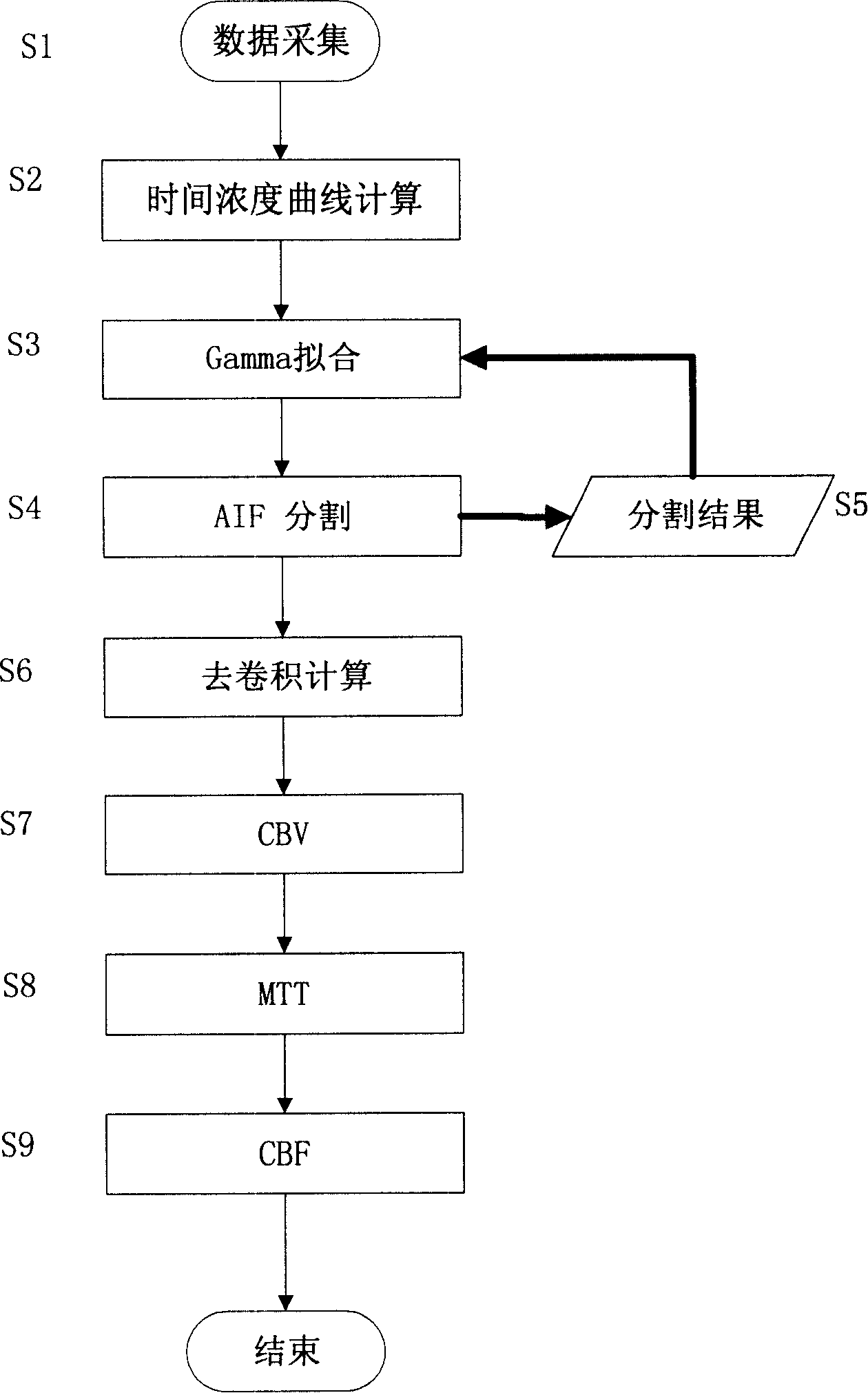
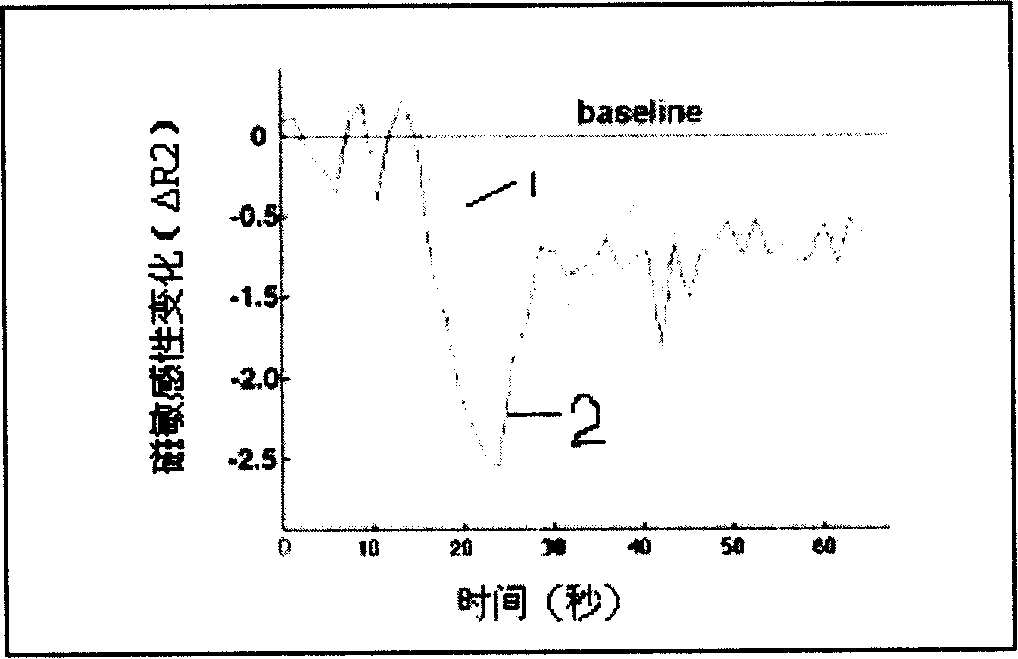
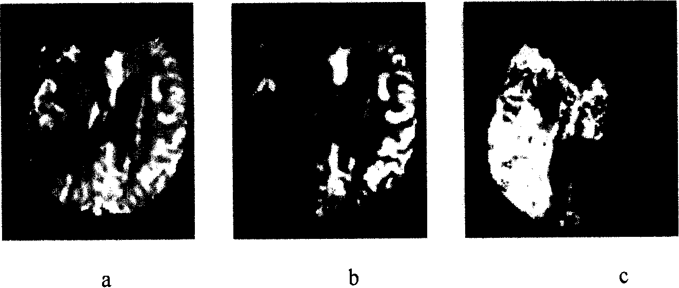
[0024] 1. Clinical observation and display of perfusion weighted imaging (PWI) images; combination: simultaneous application and analysis of PWI and ischemic penumbra area construction, and conventional magnetic resonance imaging (T1 weighted, T2 weighted, enhanced scan ) for clinical reference;
[0025] 2. Image analysis, processing and digitization methods: First, the original dynamic magnetic resonance images are collected with magnetic resonance equipment. Secondly, apply information processing means, and if necessary, use the method of absolute quantification to realize image reconstruction for the collected images;
[0026] 3. Clinical application of the digital imaging method: the proposed digital imaging method can be used clinically to make a definite diagnosis and give corresponding treatment to the early stage of acute ischemic cerebrovascular disease, and to achieve the early recovery of such patients. Purpose. According to the analysis results of this digital im...
PUM
 Abstract
Description
Claims
Application Information
 - Generate Ideas
- Intellectual Property
- Life Sciences
- Materials
- Tech Scout
- Unparalleled Data Quality
- Higher Quality Content
- 60% Fewer Hallucinations
Browse by: Latest US Patents, China's latest patents, Technical Efficacy Thesaurus, Application Domain, Technology Topic, Popular Technical Reports.
© 2025 PatSnap. All rights reserved.Legal|Privacy policy|Modern Slavery Act Transparency Statement|Sitemap|About US| Contact US: help@patsnap.com