Fluid cell equipment-microcarrier clinical diagnosis chip
A technology of flow cytometry and clinical diagnosis, applied in the field of flow cytometry-microcarrier clinical diagnosis chip, which can solve problems such as confusion, technical complexity, and affecting the affinity of biomolecules
- Summary
- Abstract
- Description
- Claims
- Application Information
AI Technical Summary
Problems solved by technology
Method used
Image
Examples
preparation example Construction
[0025] Preparation of microcarriers - biomolecules (antigens / antibodies)
[0026] The preparation of microcarrier-antigen / antibody complexes involves binding protein molecules such as antigens / antibodies to the surface of polymer particles through covalent bonds. The diameter of the polymer particles can be 1-100 microns, preferably 1-50 microns, most ideally 1-20 microns. The material of polymer particles can vary according to needs, for example, latex, silicon, resin, and various plastic polymer materials, including styrene, bromostyrene, acrylic acid, acrylic amine, methacrylate, vinyl chloride, Polyurethane and polymerizable monomers made of chlorinated styrene, ethylene acetate, etc. The covalent bonding between polymer particles and antigens / antibodies is accomplished through groups such as epoxy resin, acetaldehyde, and carbon diamine. It can also be accomplished by intermediate molecules carrying amine groups or other reactive groups such as succinyl esters. Unbound...
example 1
[0036] Example 1 Infectious Disease Detection System
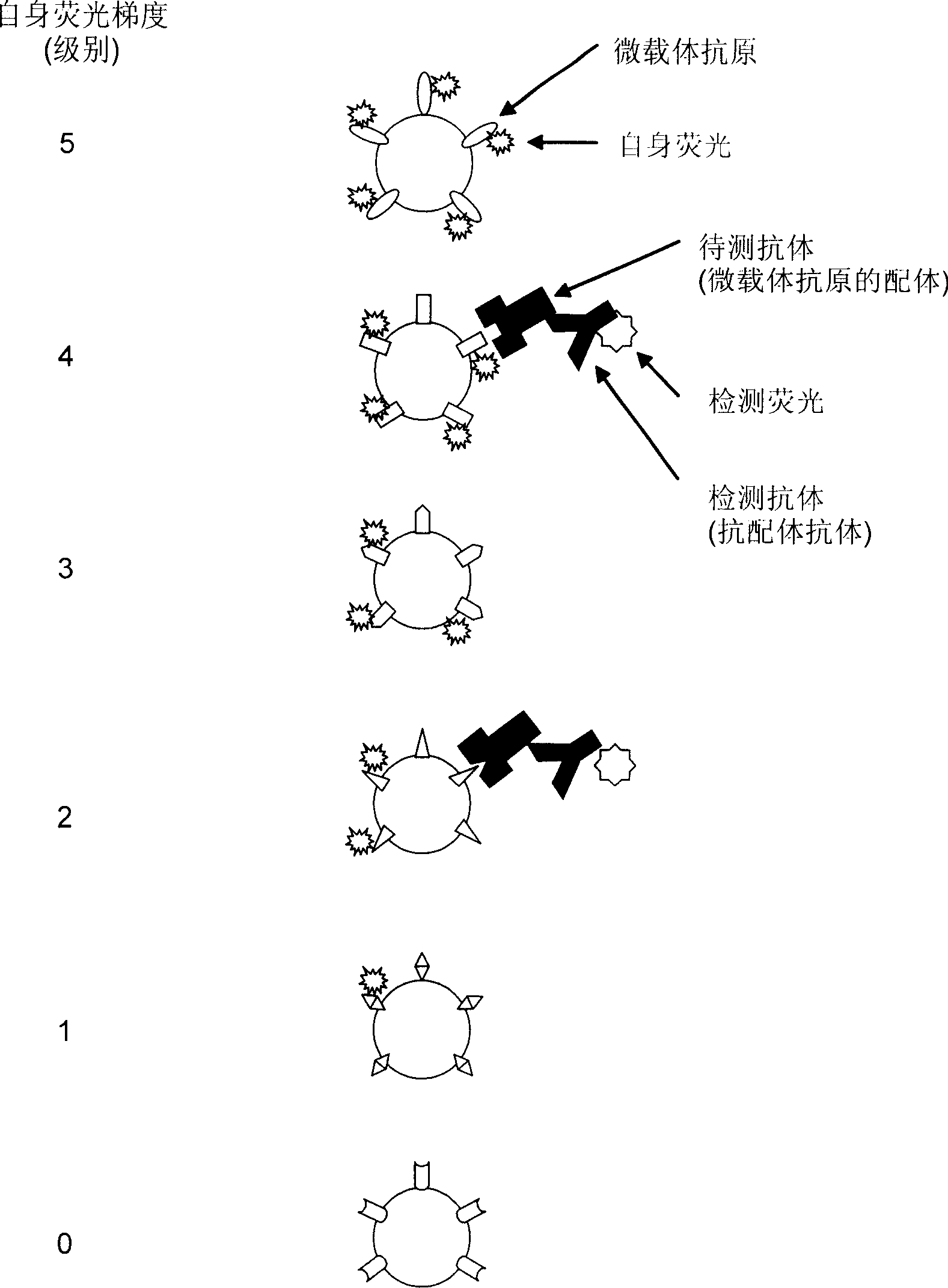
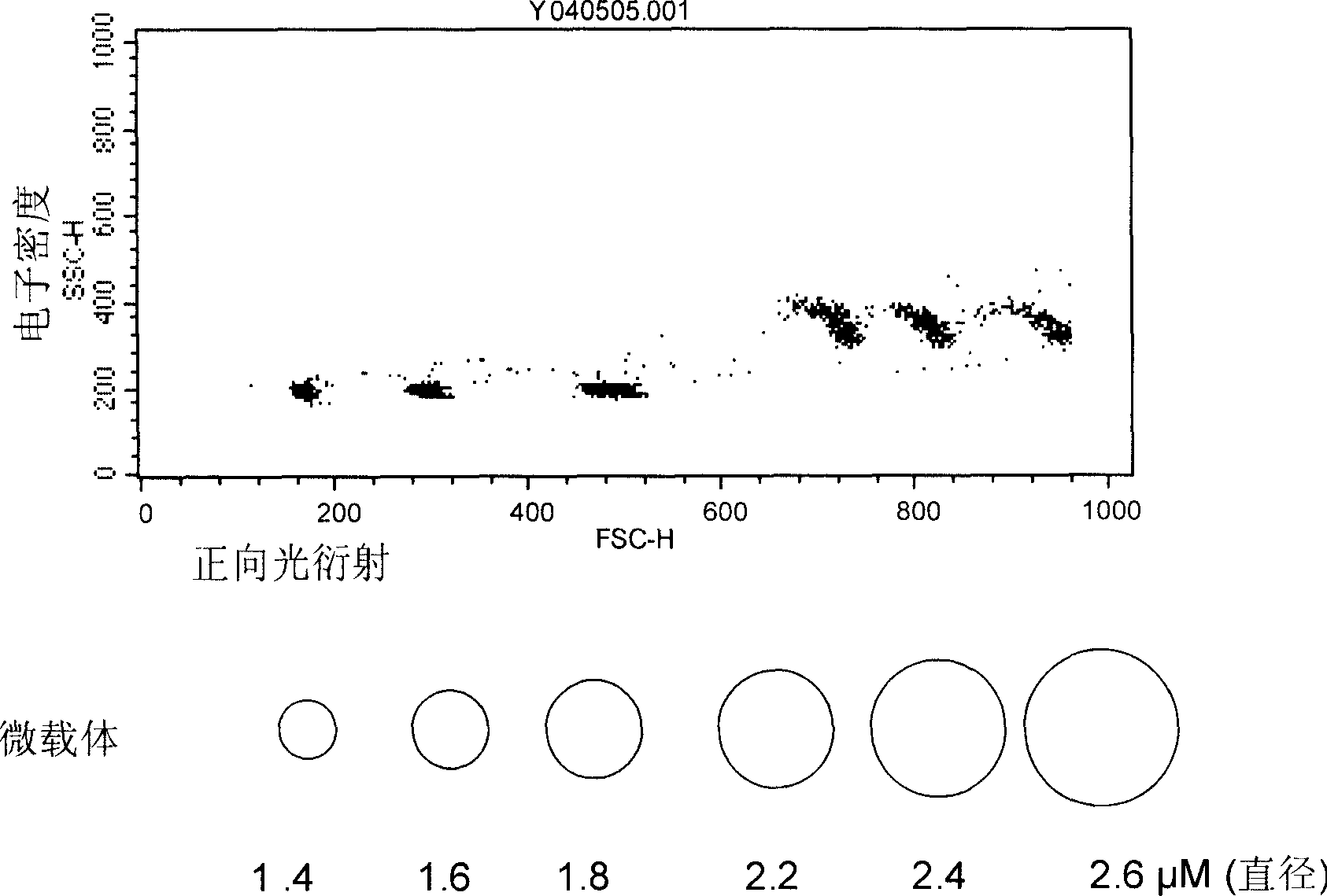
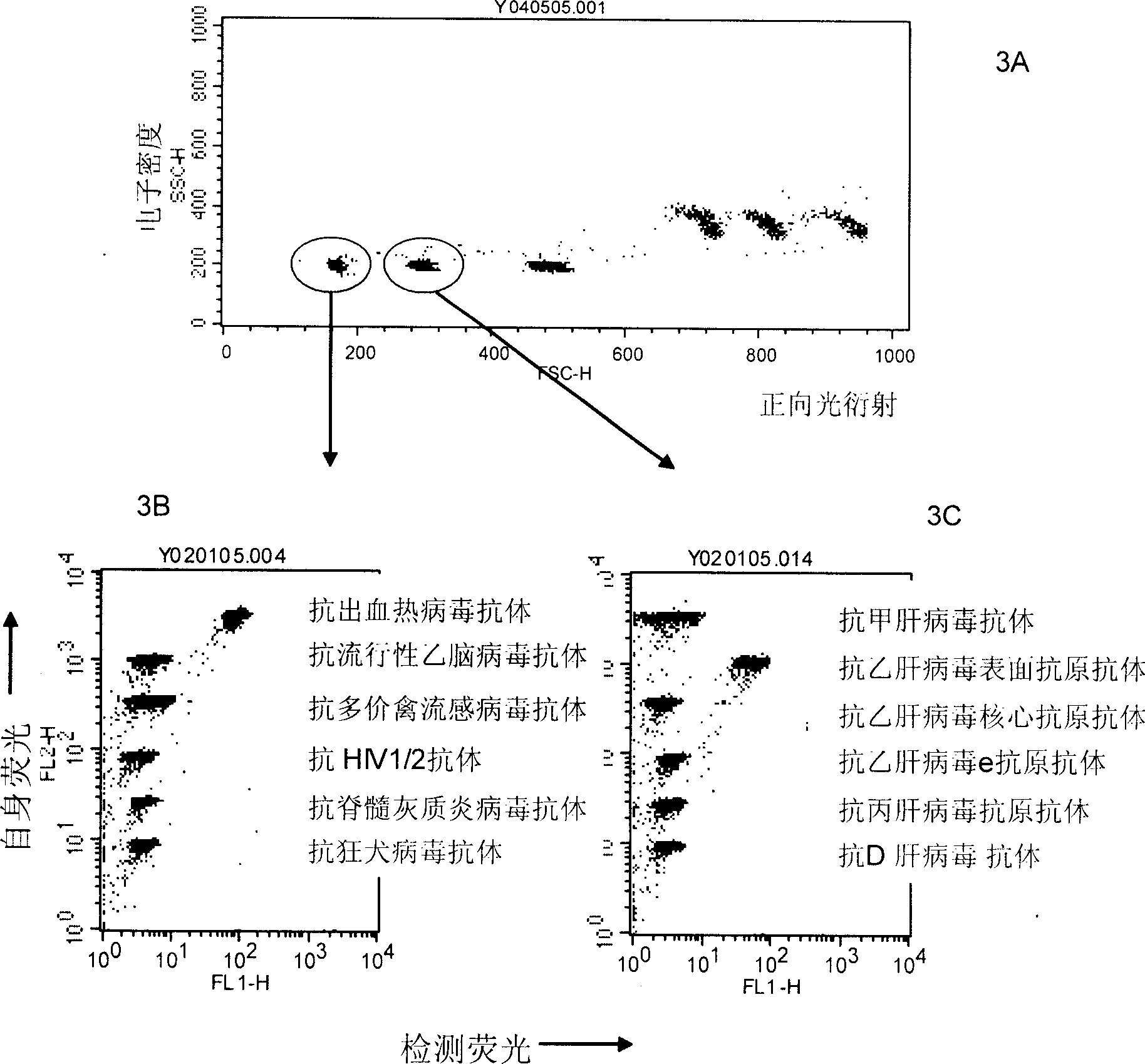
[0037] According to one of the specific embodiments of the present invention, latex particles of one diameter are respectively bound to at least 6 kinds of purified pathogen antigens or antibodies through covalent bonding. Then, the protein molecules bound to the six kinds of latex particles were labeled with different intensities of autofluorescence (such as PE from 1 μM to 10 μM) to form a fluorescence gradient (such as from 0 level to 5 level). Incubate at least one autofluorescent latex particle, or latex particle with at least one diameter, with a biological sample (such as human serum, culture fluid) to bind the ligand to be tested. The ligand to be tested bound to the latex particles was then labeled with FITC-goat anti-human IgG / IgM / IgA. The size of latex particles was determined by electron density (vertical axis) versus forward light diffraction (horizontal axis). Select latex particles with a single diameter a...
example 2
[0057] Example 2. Allergic test system
[0058] According to another specific embodiment of the present invention, latex particles of one diameter are respectively bound to six purified allergens through covalent bonding. Then six kinds of protein molecules bound to the latex particles were labeled with different intensities of autofluorescence (such as PE from 1 μM to 10 μM) to form a fluorescence gradient (such as from 0 level to 5 level). At least one autofluorescent latex particle, or at least one diameter of latex particle, is incubated with a biological sample (eg, human serum) to bind to a specific ligand, anti-allergen IgE. The anti-allergen IgE bound to the latex particles was then labeled with FITC-goat anti-human IgE. See Example 1 for flow cytometry analysis.
[0059] latex particles
(diameter)
(level)
Microcarriers - Antigen / Antibody
detection antibody
1-20μm
0
anti-human IgE
anti-huma...
PUM
 Abstract
Description
Claims
Application Information
 - R&D
- Intellectual Property
- Life Sciences
- Materials
- Tech Scout
- Unparalleled Data Quality
- Higher Quality Content
- 60% Fewer Hallucinations
Browse by: Latest US Patents, China's latest patents, Technical Efficacy Thesaurus, Application Domain, Technology Topic, Popular Technical Reports.
© 2025 PatSnap. All rights reserved.Legal|Privacy policy|Modern Slavery Act Transparency Statement|Sitemap|About US| Contact US: help@patsnap.com


