Liquid phase chip for parallel detection of autoantibodies, preparation method and application thereof

A parallel detection, autoantibody technology, applied in the field of immune technology and clinical detection, can solve problems such as inconvenience and achieve the effect of saving detection costs

Inactive Publication Date: 2006-11-22
SHANDONG MEDICAL BIO TECH RES CENT
View PDF0 Cites 16 Cited by
  • Summary
  • Abstract
  • Description
  • Claims
  • Application Information

AI Technical Summary

Problems solved by technology

[0003] According to the clinical characteristics of autoimmune diseases, which are often characterized by the emergence of a diverse autoantibody spectrum, the current ELISA detection, immunofluorescence and other single detections for existing autoantibodies can only detect one autoantibody at a time. Inconvenience and clinical diagnosis The problem to be solved by the present invention is to provide a liquid phase chip for parallel detection of autoantibodies and its preparation method and application, so as to provide a convenient and accurate clinical method for early detection, early diagnosis and early treatment of autoimmune diseases. Detection method and detection kit

Method used


Image

  • Liquid phase chip for parallel detection of autoantibodies, preparation method and application thereof
  • Liquid phase chip for parallel detection of autoantibodies, preparation method and application thereof

Examples


Embodiment 1

[0023] Example 1: Coupling of self-antigens to known numbered microspheres

[0024] 1. Select No. 28, 38, 48, 58, 68, and 78 carboxyl microspheres (Luminex Company) respectively, and oscillate the microsphere suspension with a vortex shaker for 20 seconds to mix the microspheres evenly.

[0025] 2. Take about 2×10 carboxyl microspheres of each number above. 3 Transfer each to a centrifuge tube and centrifuge at ≥8000×g for 2 min to precipitate carboxylated microspheres.

[0026] 3. Remove the supernatant and add 100μl dH 2 O, use a vortex shaker to resuspend the microspheres for 20 seconds, centrifuge at ≥8000×g for 2 minutes, and precipitate the carboxylated microspheres.

[0027]4. Remove the supernatant, add 80 μl, 100 mM, pH=6.2 sodium dihydrogen phosphate salt solution, and resuspend the washed carboxy microspheres with a vortex shaker for 20 seconds.

[0028] 5. Add 10μl, 50mg / ml Sulfo-NHS (with dH 2 O dilution), oscillate gently with a vortex shaker.

[0029] 6. Ad...

Embodiment 2

[0040] Example 2: Application of liquid phase chip for parallel detection of autoimmune diseases described in the present invention in clinical detection

[0041] (1) The technical process of using the autoantibody parallel detection liquid phase chip of the present invention to detect autoantibodies:

[0042] 1. Take out 500 microspheres conjugated to each autoantigen prepared above, mix them in equal proportions, and distribute them in 96-well plates. Each well contains 500 microspheres conjugated to various autoantibodies. Add 50 μl of serum to be tested and incubate at 37°C for 2 hours.

[0043] 2. Centrifuge at ≥8000×g for 2 minutes, remove the supernatant, add 300 μl of 1% PBS-BSA, vortex for 30 seconds, and seal in a 37°C incubator for 1 hour.

[0044] 3. Centrifuge at ≥8000×g for 2 minutes. Remove the supernatant, add 300 μl PBS-TBN, and vortex for 30 seconds. Centrifuge at ≥8000×g for 2 minutes.

[0045] 4. Repeat step 3 twice.

[0046] 5. Add 100 μl of the bioti...

Embodiment 3

[0055] Example 3: Example 1 of standard curve drawing of autoantibodies

[0056] According to the method described in Examples 1 and 2, SSA / Ro-52 (R052) was coated onto No. 38 microspheres. After coating, 1000 microspheres were added to PE-labeled mouse anti-human IgG to react for 30 minutes, and detected by Luminex100 , the fluorescence value reached 23,000, while the microspheres not coated with SSA / Ro-52 were reacted with PE-labeled mouse anti-human IgG for 30 minutes. After Luminex100 detection, the fluorescence value was only 100, indicating that our coated microspheres can meet the needs of chip development. requirements.

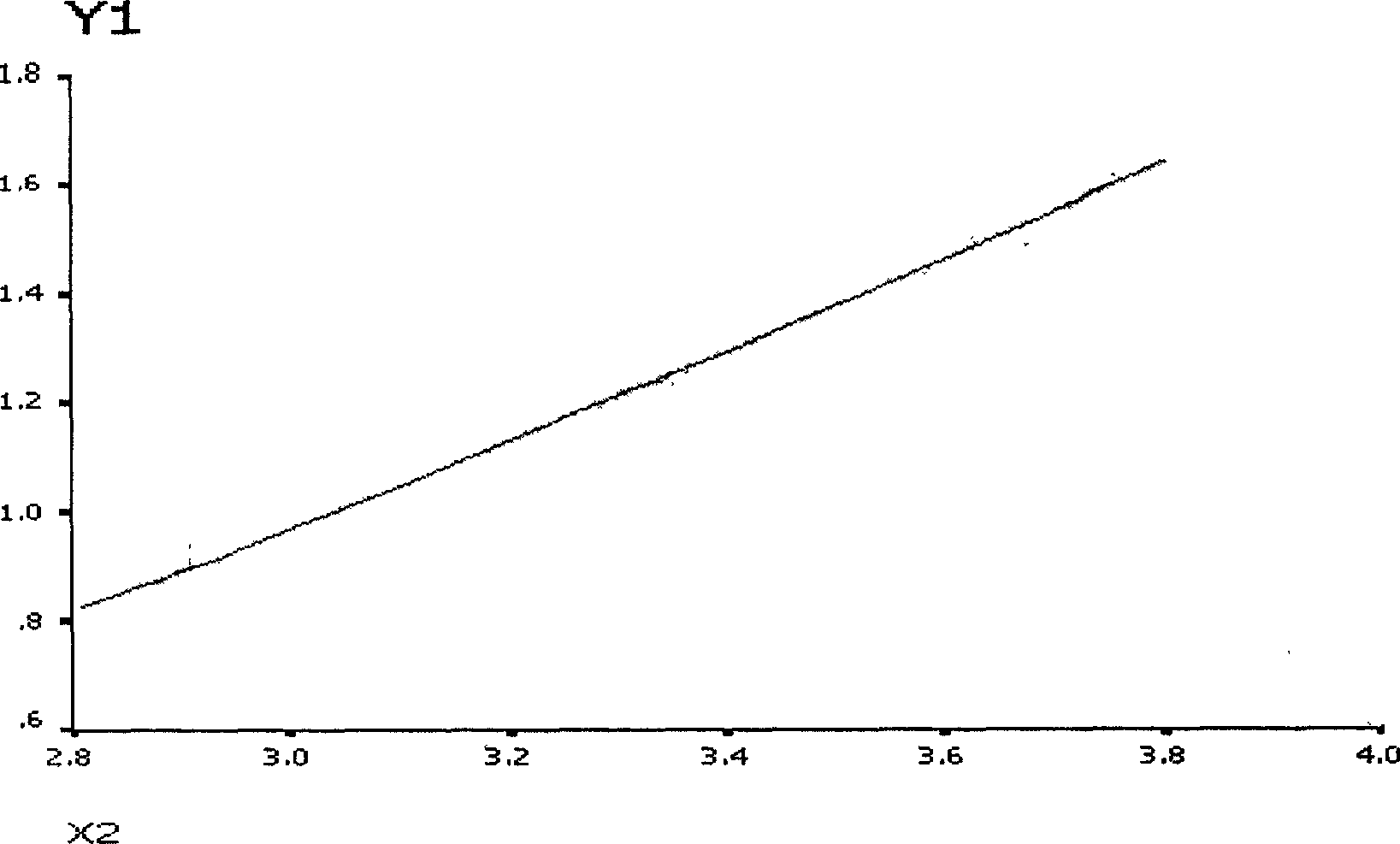
[0057] Take the standard SSA / Ro-52 antibody, dilute it according to the gradient of 0EU / ml, 6.25EU / ml, 12.5EU / ml, 25EU / ml and 50EU / ml, react according to the method described, and detect by Luminex100, the average fluorescence intensity is respectively for 261.5, 908, 1475, 4422 and 6654.5. For the standard curve obtained, see figure 1 . One case ...


PUM

PropertyMeasurementUnit
fluorescenceaaaaaaaaaa

Abstract

The related autoantibody parallel detection liquid chip comprises: the mcirosphere, self antigen to form coupling body with corresponding microsphere and use red laser to activate red fluorescence on substrate and determine self antigen type by color, and double antigen treated by biotin to combine and activate the phycoerythrin with green laser and determine fluorescent molecule quantity for indirect obtaining content.

Description

technical field [0001] The invention relates to a liquid phase chip and its preparation method and application, in particular to an autoantibody detection parallel detection liquid phase chip for autoimmune disease diagnosis and its preparation method and application, belonging to the technical field of immune technology and clinical detection . Background technique [0002] The incidence rate of autoimmune diseases in the population is 3-5%, especially the highest incidence rate among the elderly. The most common feature of these diseases is the presence of autoantibodies in the serum, and different autoantibody profiles can distinguish the various diseases in this group. In addition to the corresponding clinical symptoms, the current international diagnostic criteria for autoimmune diseases are mainly based on the detection of autoantibodies in the patient's serum. Rheumatism is difficult to diagnose in the early stage, but in the late stage of the disease, multiple orga...

Claims


Application Information

Patent Timeline
Patent Type & Authority Applications(China)
IPC IPC(8): G01N33/543G01N21/84G01N33/546
Inventor 高雪芹张华宁韩金祥徐华
Owner SHANDONG MEDICAL BIO TECH RES CENT
Why Eureka
Try Eureka