Inverse patterning process for three-dimensional multi-compartmental micro-organization of multiple cell types

a multi-compartmental, cell-type technology, applied in the field of inverse patterning process for three-dimensional multi-compartmental micro-organization of multiple cell types, can solve the problems of poor printer resolution, printing time and specific material conductivity, low cellular density of microstructures, etc., and achieve the effect of rapid cell pattern

Active Publication Date: 2018-09-11
MASSACHUSETTS INST OF TECH
View PDF6 Cites 5 Cited by
  • Summary
  • Abstract
  • Description
  • Claims
  • Application Information

AI Technical Summary

Benefits of technology

[0006]The present invention features an “inverse patterning” methodology referred to as or “Intaglio-Void / Embed-Relief Topographic (InVERT) molding.” The inverse patterning” or “InVERT” platform described herein offers numerous advantages and improvements over existing methods for patterning cells in 3D. First, the versatility of previous technologies such as laser printing, dielectrophoresis, photo-patterning, and cell “bioprinting” (see e.g., Albrecht, D. R, et al. Nat Methods 3, 369-375, (2006); Nelson, C. M., et al. Science 314, 298-300, (2006); Ungrin, M. D., et al. PLoS One 3, (2008); Du, Y, et al. Proc Natl Acad Sci USA 105, 9522-9527 (2008); Tan, W. & Desai, T. Biomaterials 25, 1355-1364, (2004); Liu Tsang, V. et al. FASEB J 21, 790-801, (2007); Akselrod, G. M. et al. Biophys J 91, 3465-3473, (2006); Mironov, V., et al. Trends Biotechno / 21, 157-161, (2003); Barron, J. A, et al. Biomed Microdevices 6, 139-147 (2004); Jakab, K, et al. Proc Natl Acad Sci USA 101,2864-2869, (2004); Tsuda, Y. et al. Biomaterials 28, 4939-4946, (2007); Mironov, V. et al. Biomaterials 30,2164-2174, (2009); and Nahmias, Y., et al. Biotechnol Bioeng 92, 129-136, (2005)) is limited by the technical parameters of the system such as poor printer resolution, printing time, and specific material conductivities.
[0008]The inverse patterning process described here yields high-resolution multi-cellular microstructures that can be patterned in distinct compartments in 3D hydrogels. This process is highly versatile. It has been shown that the technology is compatible with material systems and patterning substrates found routinely in basic biological laboratories (e.g., agarose or fibrin) as well as highly-tunable and customizable materials (e.g., polyethylene glycol) found in specialized engineering and chemistry laboratories. This process can also be used to pattern cells that have been pre-incubated to enable the formation of intercellular junctions in a given compartment or immediate trapping to produce rapid cell patterns. This process is based on cellular sedimentation and therefore invokes minimal damage to cells. Finally, this process achieves multi-compartmental patterning without the need for manual alignment of cell layers and can produce scaled tissues of clinically relevant sizes.

Problems solved by technology

Biotechnol Bioeng 92, 129-136, (2005)) is limited by the technical parameters of the system such as poor printer resolution, printing time, and specific material conductivities.
Second, to build tissues containing multiple distinctly patterned cell types, many of the existing technologies first require patterning of each cell type in a separate hydrogel layer separately followed by tedious manual alignment and merger of layers.
Third, these technologies typically rely on external physical forces such as electricity, UV light, and mechanical perturbation that have the potential to damage living cells.
Finally, most patterning technologies use “immediate trapping” of cells in a biomaterial without prior cell-cell pre-aggregation, which results in low cellular density in microstructures and interferes with the formation of intercellular junctions that are critical for normal physiologic function in many tissues.
In short, previous “top-down” engineering approaches have not achieved robust micro-scale, multi-cellular, and multi-compartmental patterning.
Alternatively, “bottom-up” approaches simply have not yet achieved the efficiency and scalability required for large-scale and high-throughput construction of engineered tissues and model systems.

Method used


Image

  • Inverse patterning process for three-dimensional multi-compartmental micro-organization of multiple cell types
  • Inverse patterning process for three-dimensional multi-compartmental micro-organization of multiple cell types
  • Inverse patterning process for three-dimensional multi-compartmental micro-organization of multiple cell types

Examples


example 1

Fabrication of Exemplary Inverse Patterned Constructs—InVERT Molding to Fabricate Engineered Tissues with Distinct Multicellular Micro-compartments

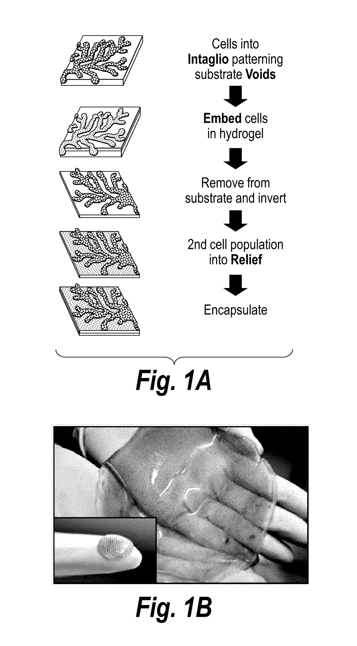
[0108]The present invention features a process by which to encapsulate multiple cell types with distinct organization into a 3D hydrogel. (FIG. 1a). In particular, scalable, versatile, and rapid 3D multi-compartmental cellular patterning was achieved using an InVERT molding protocol (FIG. 1a). Topographic substrates were first produced containing microscale features and replica molded these substrates using poly(dimethylsiloxane) (PDMS) to create topographic ‘intaglio’ cell capture substrates with recessed ‘voids’. In this example, cells were first isolated in the micro-scale features of polydimethyl siloxane (PDMS) cell-capture substrate either in media or in a pre-polymer material. Cells patterned in media were then incubated overnight to allow formation of cell-cell junctions, e.g., cadherin and adhesion junctions, and then encapsulate...

example 2

Characterization of Multi-Compartmental Cellular Microorganization; High-fidelity Microorganization of Multiple Cell Types Across Distinct Compartments

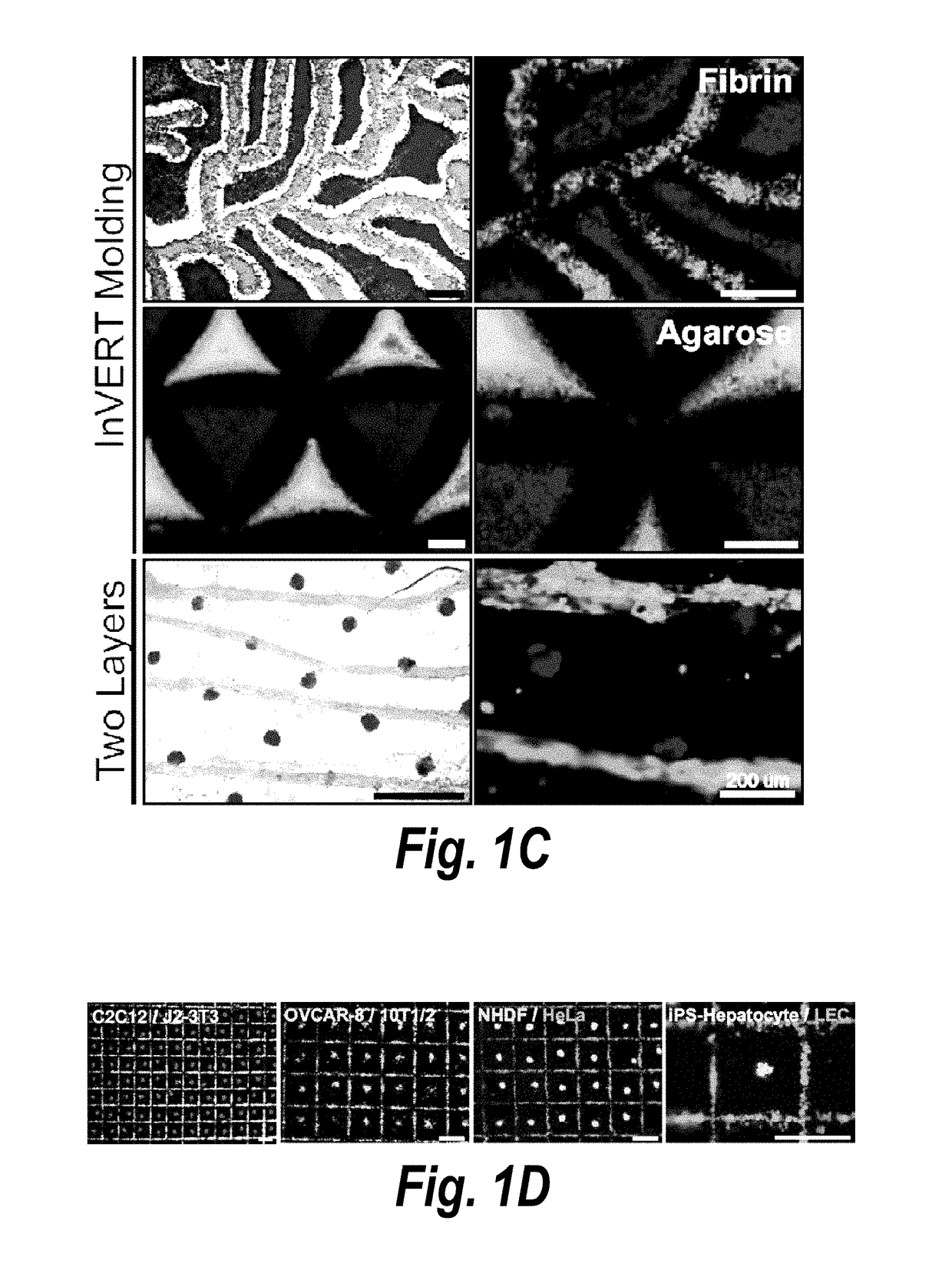
[0118]Existing scalable technologies do not allow interrogation of the functional impact of placement of multiple compartments in engineered tissues on the parenchyma. Endothelial cells of the vasculature modulate parenchymal tissue functions in both health and disease (Ding, B. S. et al. Nature 468, 310-315 (2010); Ding, B. S. et al. Cell 147, 539-553 (2011); Franses, J. W., et al. Sci Transl Med 3, 66ra65 (2011); and Aird, W. C. Pharmacol Rep 60, 139-143 (2008)) and play an ultimately non-dispensable role in oxygen and nutrient delivery. It was therefore desired to demonstrate the power of the InVERT molding technique by controlling the micro-scale patterning of distinct ‘parenchymal’ and ‘vascular’ compartments in 3D engineered liver tissue. Primary rat hepatocytes were seeded in microwell patterning substrates, incubated overnight...

example 3

Hierarchical Modulation of Microstructure and Multi-cellular Composition within a Given Compartment

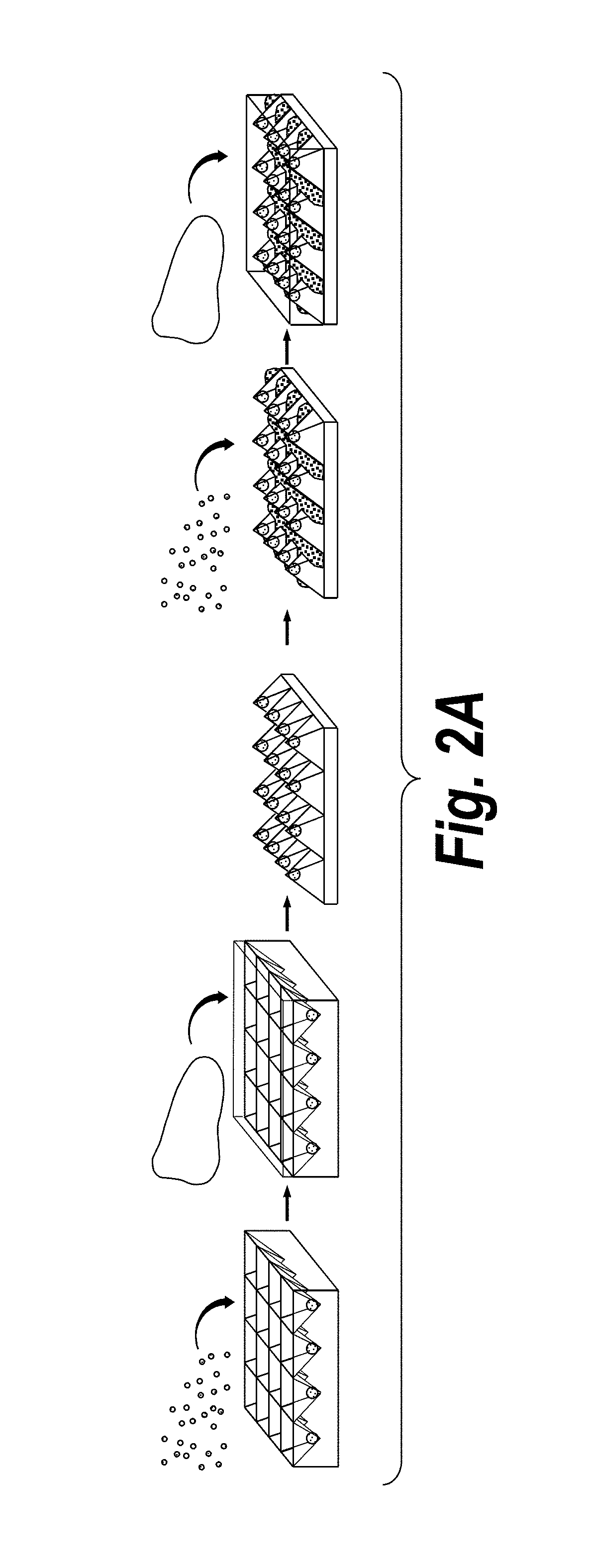
[0120]Complex tissues are hierarchically organized across multiple length-scales and contain numerous cell types. In particular, such complex tissues are hierarchically organized such that each tissue compartment is further organized at both the microstructural and multicellular level. Thus, it was next examined whether additionally fine-tuning of the microstructure and multi-cellular composition within a tissue given compartment could be accomplished using InVERT molding. Specifically, we refined the hepatic compartment produced during the initial Intaglio-Void-Embed portion of the InVERT molding protocol without adding a second cell population (‘relief’ phase). To test whether the microstructure of aggregates in the hepatic compartment could be reproducibly modulated, primary rat hepatocytes were seeded in microwell patterning substrates at densities of 10-500 cells per microwell, in...


PUM

PropertyMeasurementUnit
diameteraaaaaaaaaa
diameteraaaaaaaaaa
diameteraaaaaaaaaa

Abstract

The invention features an “inverse patterning” or “Intaglio-Void / Embed-Relief Topographic (In VERT) molding” manufacturing process for generating high-resolution three-dimensional (3D) multi-cellular microstructures in distinct cellular compartments of a single hydrogel. The platform has general utility in the development of engineered tissues for human therapies, drug testing, and disease models. Additionally, the platform can serve as a model system for studying 3D cell-cell interactions in fields as diverse as stem cell biology to the development of cancer therapeutics.

Description

CROSS-REFERENCE TO RELATED APPLICATIONS[0001]This application is a 35 U.S.C. 371 national stage filing of International Application PCT / US2013 / 028345, filed 28 Feb. 2013, which claims priority to U.S. Patent Application No. 61 / 604,841, filed 29 Feb. 2012 and U.S. Patent Application No. 61 / 619,074, filed 2 Apr. 2012. The contents of the aforementioned are hereby incorporated by reference.STATEMENT OF FEDERALLY SPONSORED RESEARCH[0002]This invention was made with government support under grant nos. EB008396 and DK56966 awarded by the National Institutes of Health (NIH). The government has certain rights in the invention.BACKGROUND[0003]Cells sense and respond to cues in local their environment, such as signals produced by neighboring cells as well as the chemistry and mechanics of the surrounding matrix. The spatial organization of cells within tissues, or tissue “architecture”, defines the cell-cell contacts and paracrine signaling gradients thought to ultimately drive tissue functio...

Claims


Application Information

Patent Timeline
Patent Type & Authority Patents(United States)
IPC IPC(8): C12N5/00C12N11/04G01N33/50A61K35/12C12N5/071C12N11/10C12N11/08
CPCC12N11/04A61K35/12C12N5/0012C12N5/0062C12N11/08C12N11/10G01N33/5005C12N5/0671C12N2535/10C12N2533/76C12N11/089C12N11/098
Inventor BHATIA, SANGEETA N.STEVENS, KELLY R.
Owner MASSACHUSETTS INST OF TECH
Why Eureka
Try Eureka