
Ionizing irradiation sterilization of bacterial minicell-based biopharmaceuticals and methods of use
a technology of biopharmaceuticals and minicells, which is applied in the field of compositions and methods, can solve the problems of non-living minicells, non-infectious, and inability to replicate, and achieve the effect of ensuring consistent sterility using a validated sterilization method and not living minicells
- Summary
- Abstract
- Description
- Claims
- Application Information
AI Technical Summary
Benefits of technology
Problems solved by technology
Method used
Image

Examples
example 1
No Change in VAX-IP (VAX014) Minicell Properties in a Frozen Suspension Following Exposure to Ionizing Gamma Irradiation at Doses of 5, 15 and 25 kGy
[0164]This example shows that there was no change in VAX-IP (VAX014) minicell properties following exposure to ionizing gamma irradiation in a frozen suspension at doses of 5, 15 and 25 kGy.
[0165]VAX014 minicells (contains both Invasin and Perfringolysin O) were made from parental strain VAX20B9, a recombinant Escherichia coli K-12 minicell-producing strain harboring the L-rhamnose-inducible bacterial expression plasmid, pVX-336 (containing a transcriptional fusion between Invasin and Perfringolysin O under control of the pRHABAD promoter). One vial of VAX20B9 research cell bank was thawed at room temperature and the entire vial contents used to inoculate a 1 L starter culture in Soytone-based MSB-NaCl media containing kanamycin (50 μg / mL), diaminopimelic acid (100 μg / mL), lysine (55 μg / mL), and 0.2% D-glucose. The starter culture was g...
example 2
No Change in VAX-IP (VAX014) Minicell Properties in a Frozen Lyophile Following Exposure to Ionizing Gamma Irradiation at Doses of 5, 15 and 25 kGy
[0168]This example shows that there was no change in VAX-IP (VAX014) minicell properties following exposure to ionizing gamma irradiation in a frozen lyophile at doses of 5, 15 and 25 kGy.
[0169]VAX014 minicells (contains both Invasin and Perfringolysin O) are made from parental strain VAX20B9, a recombinant Escherichia coli K-12 minicell-producing strain harboring the L-rhamnose-inducible bacterial expression plasmid, pVX-336 (contains a transcriptional fusion between Invasin and Perfringolysin O under control of the pRHABAD promoter). One vial of VAX20B9 research cell bank was thawed at room temperature and the entire vial contents used to inoculate a 1 L starter culture in Soytone-based MSB-NaCl media containing kanamycin (50 μg / mL), diaminopimelic acid (100 μg / mL), lysine (55 μg / mL), and 0.2% D-glucose. The starter culture was grown fo...
example 3
Studies of Minicell-Containing Compositions Following Exposure to Gamma Irradiation
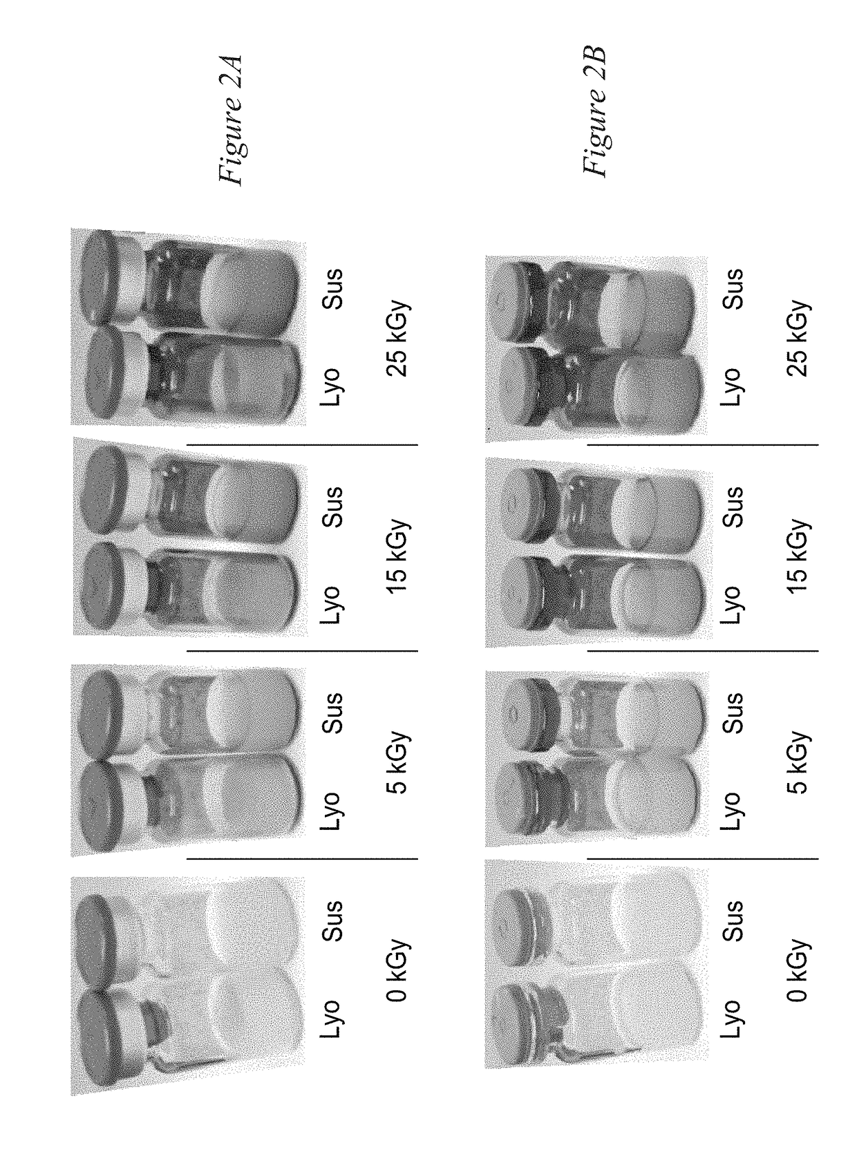
[0172]Clear vials were selected for the purpose of ensuring and demonstrating that increasing levels of ionizing gamma irradiation were administered. Vials containing minicells were exposed to gamma irradiation at 5 kGy, 15 kGy or 25 kGy, and compared to non-irradiated controls (i.e., 0 kGy). FIGS. 2A-B show the results of the initial visual inspection, held at the vial level, post-irradiation. It was found that vials were darkened with increasing irradiation dose level, yet there was no visual loss of product (including volume and drug substance). FIG. 2A shows sealed vials and FIG. 2B shows vials where the seals have been removed and the stoppers remain intact upon visual inspection. No macroscopic abnormalities were observed in the over-seal or stopper.
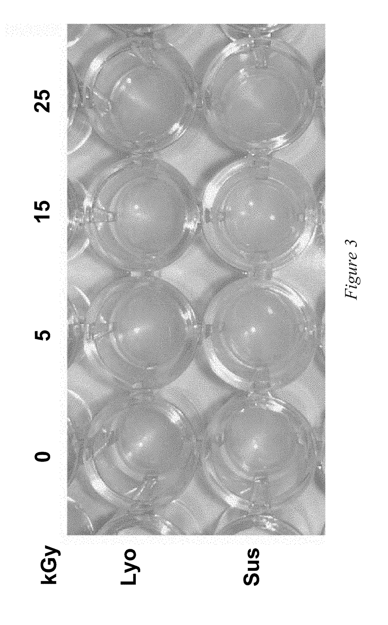
[0173]FIG. 3 further shows that there was no change in appearance (including color, consistency, and opaqueness) per secondary visual inspection of ...
PUM
| Property | Measurement | Unit |
|---|---|---|
| diameter | aaaaa | aaaaa |
| molar ratio | aaaaa | aaaaa |
| temperature | aaaaa | aaaaa |
Abstract
Description
Claims
Application Information