Kits and methods for evaluating, selecting and characterizing tissue culture models using micro-RNA profiles
a tissue culture model and microrna technology, applied in biostatistics, biochemistry apparatus and processes, instruments, etc., can solve the problems of not knowing whether the in vitro 3d models produced by different methods, molecular similar or identical, and not model the stromal-epithelial interaction. , to achieve the effect of negative regulation of apoptosis
- Summary
- Abstract
- Description
- Claims
- Application Information
AI Technical Summary
Benefits of technology
Problems solved by technology
Method used
Image
Examples
example 1
3D Tumor Tissue Models
[0109]Tumor spheroids and Tumor histoids (TH / 3DH) are produced in either one of the two 3D culture modes: 1. low shear, rotating suspension culture or 2. hanging drop (HD) cultures in 96 or 384 well Perfecta3D™ Hanging Drop Plates. TH are cultured in two stages: a. generation of fibroblast spheroids using normal newborn foreskin fibroblasts, b. addition of tumor cells (various ATCC lines) to the preformed spheroids.
[0110]Bioreactor Culture Procedure: (1).
[0111]120.0×10⁶ fibroblasts in 60 ml of culture medium are introduced into the culture chamber and rotated at approximately 6 rpm for five hours during which they form spheroids. (2). Tumor cells in 4 ml medium are added to completely fill the chamber and rotation is continued for the duration of culture (approximately 10 days).
[0112]Bioreactor Cultures:
[0113]Cancer cells coat and invade the fibroblast core producing extracellular matrix during culture. One 64 ml culture typically yields approximately 600 indiv...
example 2
Gene Expression Microarray Analysis
[0114]Cells of normal foreskin fibroblast (NF), breast cancer line UACC-893 (UA) and prostate cancer line DU-145 (DU) were cultured in 2D and 3D bioreactor. 2D cultures were harvested at confluence. Cells of 3D spheroids (single cell type) and histoids, with fibroblast core and tumor cells UACC-893 or DU-145 (FIGS. 3b and 3c) were cultured in bioreactor. Spheroids were harvested on day 4 and histoids on day 10 and cryopreserved. For miRNA and gene expression analysis cryopreserved samples were shipped on dry ice to Ambry Genetics, CA, a CLIA service provider.
[0115]Total RNA was isolated according to Ambion mirVana miRNA Isolation Kit protocol. RNA quality and concentration was determined on NanoDrop spectrophotometer and bioanalyzer. Labeling reactions were carried out using Agilent miRNA two part Complete Labeling and Hyb Kit (Version 2.1) with 100 ng total RNA input. Samples were placed on human 15K miRNA array V3 with a layout 1 human 8×15K miRN...
example 3
of miRNA Microarray Data
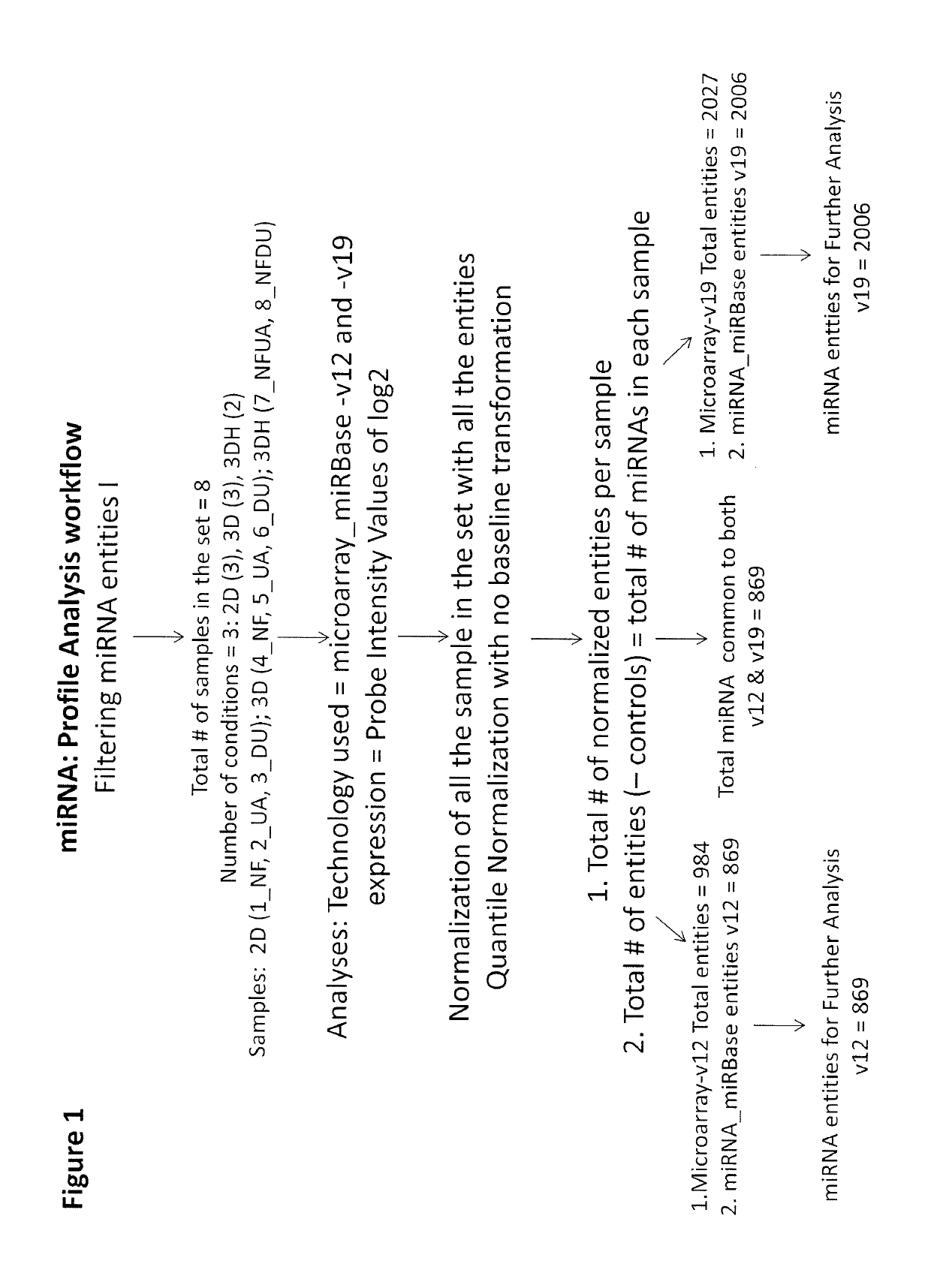
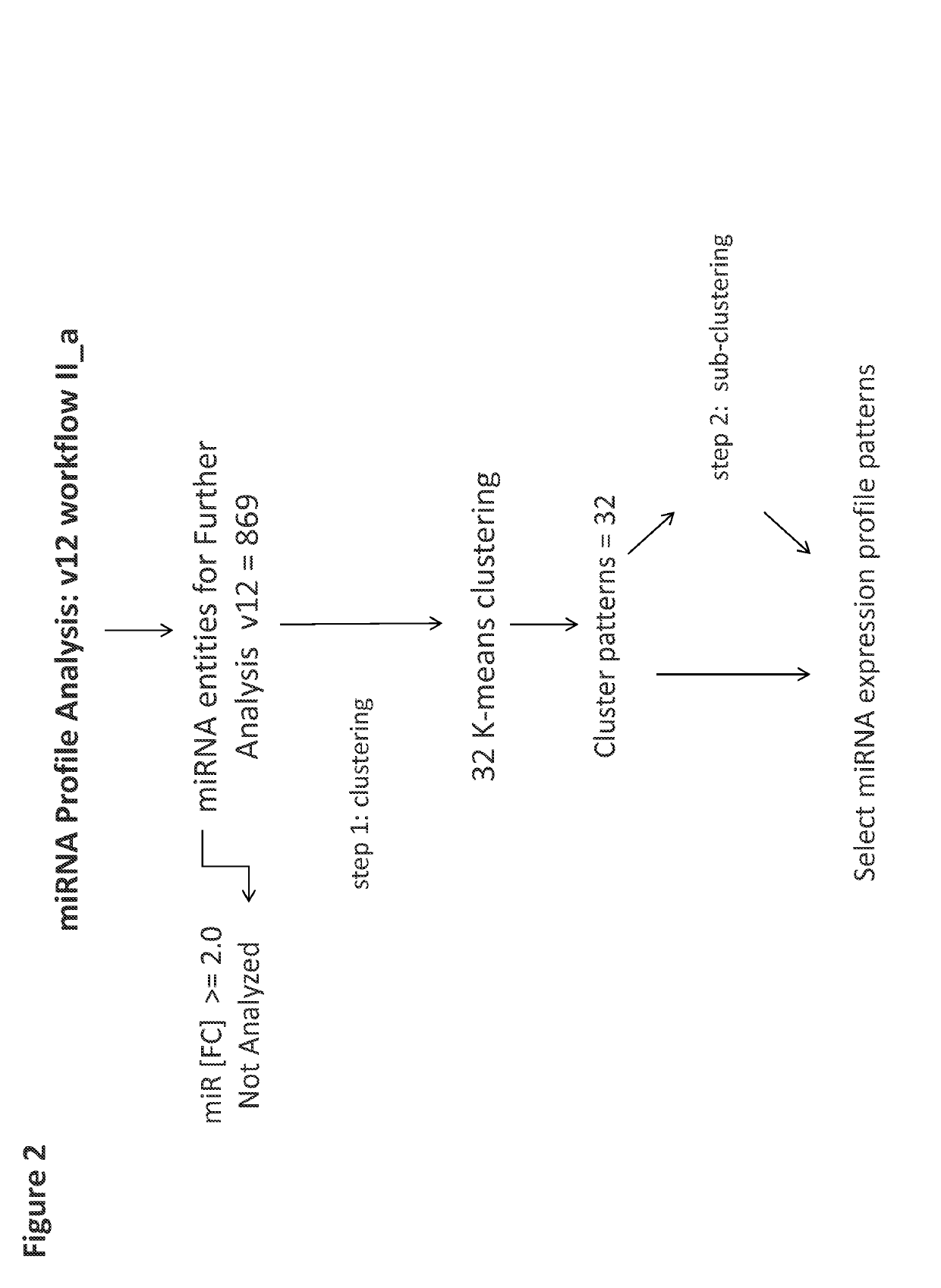
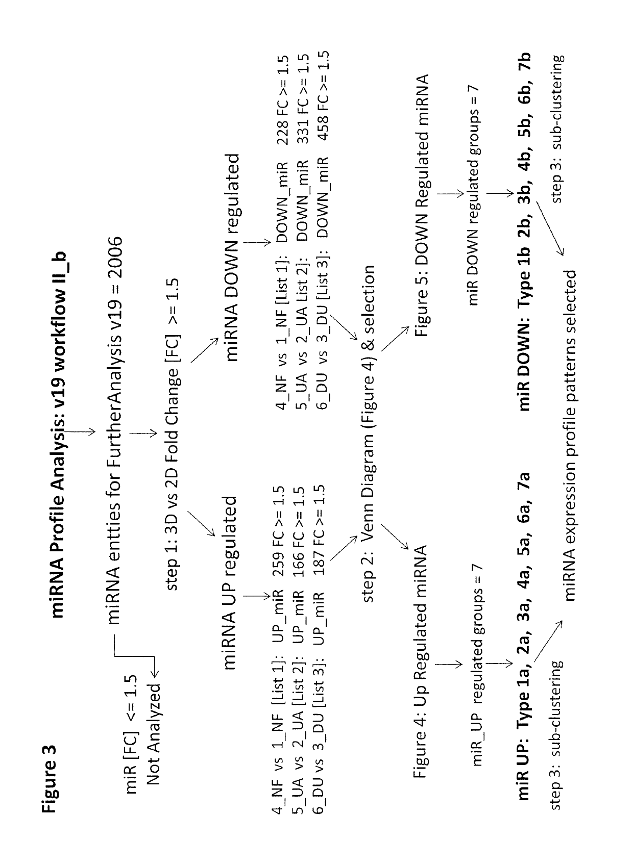
[0116]A set of 8 samples was applied to v12 or v19 miRNA microarrays ([1]-NF2D, [4]-NF3D (Normal Fibroblast), [2]-UA2D, [5]-UA3D (breast cancer), [3]-DU2D, [6]-DU3D (prostate cancer), [7]-NFUA3DH, [8]-NFDU3DH, wherein 2D designates monolayer culture, 3D designates 3D spheroid culture and 3DH designates 3D histoid culture). The raw data from (probe signal intensity values) was transformed into log 2 miRNA Gene Signal Intensity Values. Subsequently all miRNA entities across all samples in v12 (984 entities) and v19 (2027 entities) normalized by quantile normalization with no baseline transformation. At the next step All controls and all viral microRNA from normalized data set were filtered with search and Venn diagram functions. After this step: 869 miRNA entities (v12) or 2006 miRNA entities (v19) were taken forward to the next step of analysis (FIG. 1). Subsequently v12 set was subjected to 32 K-means clustering (FIG. 2). The v19 set on the other hand was sub...
PUM
| Property | Measurement | Unit |
|---|---|---|
| time | aaaaa | aaaaa |
| heat map | aaaaa | aaaaa |
| heat maps | aaaaa | aaaaa |
Abstract
Description
Claims
Application Information
 - R&D
- Intellectual Property
- Life Sciences
- Materials
- Tech Scout
- Unparalleled Data Quality
- Higher Quality Content
- 60% Fewer Hallucinations
Browse by: Latest US Patents, China's latest patents, Technical Efficacy Thesaurus, Application Domain, Technology Topic, Popular Technical Reports.
© 2025 PatSnap. All rights reserved.Legal|Privacy policy|Modern Slavery Act Transparency Statement|Sitemap|About US| Contact US: help@patsnap.com


