Methods for delivery of heart valve devices using intravascular ultrasound imaging

a heart valve and ultrasound imaging technology, applied in the field of system and method of delivering implantable medical devices, can solve the problems of excessive dilatation of the annulus of the mitral heart valve, serious problems of the heart valve incompetence, and the expansion and weakening of the chambers of the heart, so as to reduce the duration of the procedure and reduce the effect of eliminating the problem of complications

Active Publication Date: 2019-07-02
BOSTON SCI SCIMED INC
View PDF487 Cites 28 Cited by
  • Summary
  • Abstract
  • Description
  • Claims
  • Application Information

AI Technical Summary

Benefits of technology

[0010]The development relates to the implant and delivery systems, and associated methods of use of each. The implant is a device capable of extending out to the dilated annulus of a heart valve, engaging the tissue of the heart valve annulus, and gathering or cinching it in to a smaller diameter. The implant includes a tubular frame with moveable struts, where pairs of adjacent struts form apexes. The apexes have collars at least partially surrounded the apex. After engaging heart valve annulus tissue with the implant, the collars can be moved down the apex to decrease the angle between the adjacent struts, causing the tubular frame to contract in width. This pulls the tissue of the heart valve annulus closer together. In some embodiments, the device has a ring-like member formed of a shape memory material that can be expanded, e.g. forcibly expanded, to engage and penetrate the tissue of the heart valve annulus. When the force is removed, the device moves, e.g. pulls, the tissue of the heart valve annulus tissue closer together. The implant thus reconfigures. the valve annulus down to a smaller diameter, reducing and / or eliminating problems associate with the valve, such as regurgitation.

Problems solved by technology

Heart valve incompetency is a serious problem.
For example, heart disease can cause the chambers of the heart to expand and weaken.
Consequently, the annulus of the mitral heart valve dilates excessively.
In this state of dilation, valve leaflets no longer effectively close, or coapt, during systolic contraction.
This procedure is invasive as it is performed open chest and is also time consuming.
In open heart surgery, the patient is put on cardiopulmonary bypass with its associated risks of morbidity and mortality due to stroke, thrombosis, heart attack and extended recovery time.

Method used


Image

  • Methods for delivery of heart valve devices using intravascular ultrasound imaging
  • Methods for delivery of heart valve devices using intravascular ultrasound imaging
  • Methods for delivery of heart valve devices using intravascular ultrasound imaging

Examples


Embodiment Construction

,” one will understand how the features of the embodiments described herein provide advantages over existing systems, devices and methods.

[0008]The following disclosure describes non-limiting examples of some embodiments. For instance, other embodiments of the disclosed systems and methods may or may not include the features described herein. Moreover, disclosed advantages and benefits can apply only to certain embodiments of the invention and should not be used to limit the disclosure.

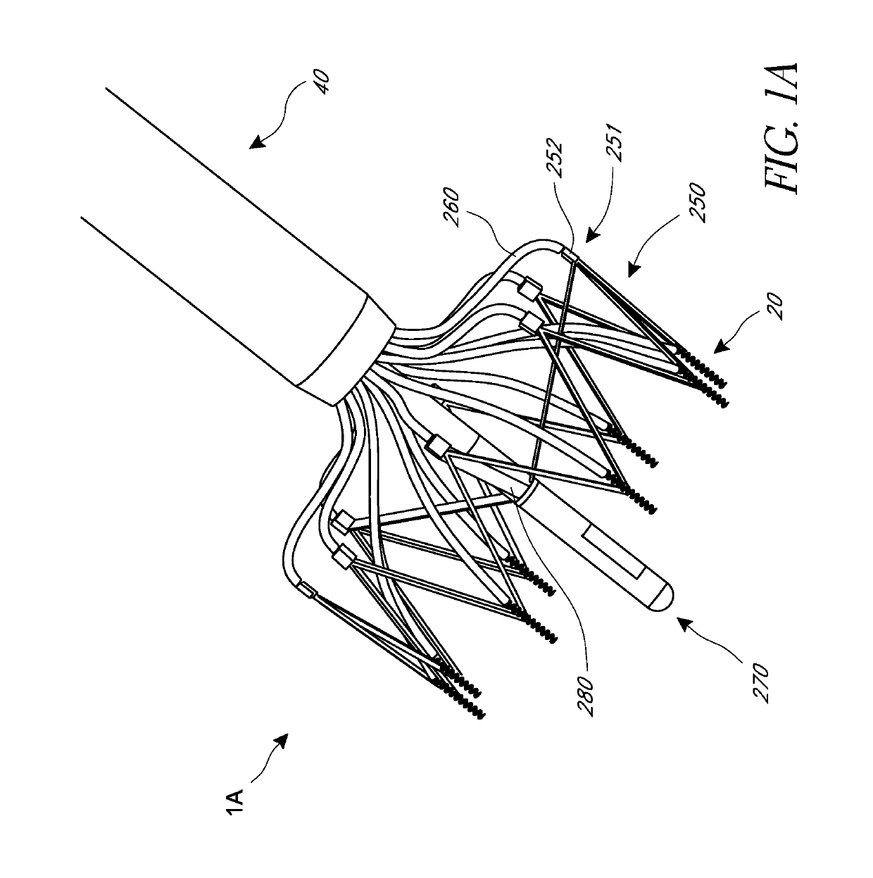
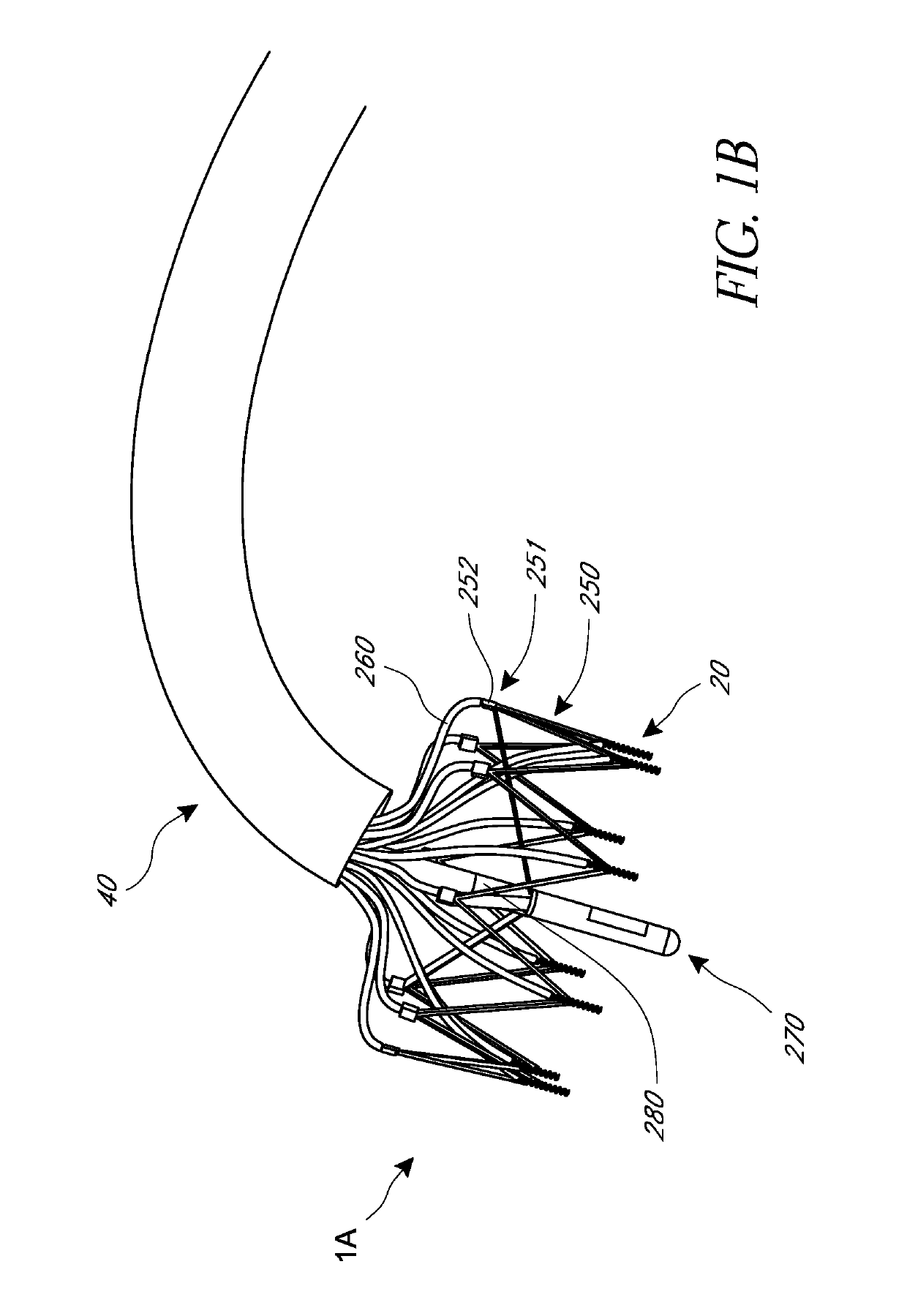
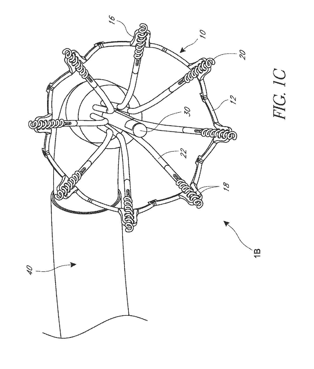
[0009]Systems and methods of delivering a heart valve implant using ultrasound imaging are described. The implant is intended to be delivered in a minimally invasive percutaneous manner, such as transfemorally, transeptally, or transapically. The implant may instead be implanted surgically, in that it should reduce the duration of the procedure and, more particularly, the duration that the patient is on bypass. Furthermore, it should be recognized that the development can be directed to mitral valve o...


PUM


Abstract

Methods related to delivery of various heart valve implants are described. The implant may be delivered using an ultrasound imaging delivery system. The ultrasound imaging delivery system may be used to deliver a variety of different devices, including mitral valve reshaping devices, mitral valve replacement valves, and others. A deployment catheter carrying an implant having a tissue anchor is advanced to a deployment site in a heart. An imaging element is positioned adjacent the implant and a relationship between the tissue anchor and an anatomical landmark in the heart is visualized. The implant is then attached by driving the tissue anchor into tissue in the heart.

Description

INCORPORATION BY REFERENCE TO ANY PRIORITY APPLICATIONS[0001]Any and all applications for which a foreign or domestic priority claim is identified in the Application Data Sheet as filed with the present application are hereby incorporated by reference under 37 CFR 1.57. For example, this application claims the benefit of priority under 35 U.S.C. § 119(e) of U.S. Provisional Application No. 62 / 234,592 entitled “HEART VALVE DELIVERY SYSTEM WITH INTRAVASCULAR ULTRASOUND IMAGING CAPABILITY” and filed on Sep. 29, 2015, the entire disclosure of which is incorporated herein by reference for all purposes and forms a part of this specification.BACKGROUNDField[0002]In general, features related to systems and methods of delivering implantable medical devices are described. For example, delivery and positioning systems and methods for implanting various devices in a heart valve, for example to treat cardiac valve insufficiency, are described.Description of the Related Art[0003]Heart valve incom...

Claims


Application Information

Patent Timeline
Patent Type & Authority Patents(United States)
IPC IPC(8): A61F2/24A61B17/064A61B8/08A61B8/12A61B8/00
CPCA61F2/2427A61B8/085A61B8/0841A61B8/0883A61B8/12A61B17/064A61F2/2412A61F2/2466A61B8/445A61B2017/0649A61B2017/0647A61B17/068A61F2/2418A61B2017/00309A61B2017/00314A61B2017/00327
Inventor LASHINSKI, RANDALLKRISTOFFERSEN, KRISTIANRUST, MATTHEWGLENN, RICHARDLEE, MICHAELMACAULAY, PATRICK
Owner BOSTON SCI SCIMED INC
Why Eureka
Try Eureka