Echo-opaque urethral catheter

a urethral catheter and echo-opaque technology, applied in the field of urethral catheter identification and treatment devices, can solve the problems of increasing the morbidity and/or intra-operative mortality of patients, destroying tumors, and unable to achieve desirable surgery for prostate removal, etc., to achieve the effect of facilitating the precise placement of treatment elements

Active Publication Date: 2020-05-05
BEST MEDICAL INT
View PDF24 Cites 5 Cited by
  • Summary
  • Abstract
  • Description
  • Claims
  • Application Information

AI Technical Summary

Benefits of technology

[0013]The present invention relates to a device and method of identifying a patient's urethra in real time, in order to facilitate the precise placement of a treatment element within the prostate in a desired location relative to the urethra. In particular, the present urethra identification device comprises a flexible elongated catheter with a primary lumen and a tip on its structured to be inserted into a patient's urethra into which is in fluid communication with a urinary bladder of the patient. Further, the elongate catheter will preferably have a sufficient length such that a tip of the catheter will actually extend into the urinary bladder of the patient. In a preferred embodiment, a marker is provided generally at the proximal tip of the elongate catheter. The tip marker may be made of hyper-echogenic and radio-opaque materials and may take a special form and shape, so that it would accurately indicate the position of the proximal end of the echo-opaque catheter during the device insertion and treating elements implantation. Additionally, a tip balloon may be provided near the proximal end of the echo-opaque catheter. This balloon is structured to be inflated once introduced into a patient's urinary bladder and thus secure the echo-opaque catheter within the patient.
[0014]The Echo-opaque catheter is structured to be at least partially echogenic and radio-opaque on its proximal portion so that an image device such as an X-ray Machine or an ultrasound device / screen, can image / see it, such as by X-ray imaging or ultrasound imaging, respectively. In a desired embodiment, the entire echo-opaque catheter is echogenic and radio-opaque, so an image of the portion of the urethra that is surrounded by a prostate can surely be seen on an image device. This design eliminates the guesswork associated with properly locating an image-generating portion of a catheter as described in the prior art.
[0019]As a result of utilizing either type of image devices, a practitioner is able to effectively identify the urethra, as well as viewing the prostate before and during introduction of the treatment elements. Moreover, the process can generate a meaningful, manageable and standardized display for an extended period of time as needed by the practitioner.
[0020]From the preceding, it is seen that the present echo-opaque urethral catheter may be desirably utilized within a method of identifying a patient's urethral anatomic course, in real time for the precise placement of a treatment element into the patient's prostate. In particular, the method desirably includes an initial step of producing a urethral catheter that is elongated, flexible and with enhanced echogenicity and radio-opaque properties. Then, an imaging probe is operatively positioned relative to the treatment site of the prostate and the urethra. This imaging device will ultimately be activated so as to obtain a real time image of the treatment site of the prostate, such as on an associated monitor, which may be viewed by a practitioner. An echo-opaque urethral catheter would be inserted into the urethra until it reaches the urinary bladder. In one embodiment, the tip of the catheter is indicated by a tip marker, which is made with materials of enhanced echogenic and radio-opaque properties. With the echo-opaque urethral catheter appropriately positioned within the urethra of the patient with the tip marker placed within the bladder or at the bladder neck, the urethra can be thereafter identified during placement of the treatment element, and proper positioning of the treatment element relative to the urethra can be ensured. The method may further include a tip balloon. The tip balloon is located near the proximal end of the catheter and which is in fluid communication with the catheter. This tip balloon is inflated once the tip of the catheter reaches the urethral neck of the patient's bladder and therefore secures the catheter in position, avoiding any movement during the implantation of treatment elements.

Problems solved by technology

Prostate cancer is potentially aggressive and is the second leading cause of cancer deaths among men in the United States.
However, in many circumstances surgery for the removal of a prostate may not be desirable for a variety of reasons.
Among these are the postoperative risks of urinary incontinence and erectile dysfunction, and co-morbid medical conditions, which may increase a patient's morbidity and / or intra-operative mortality.
In this regard, the one or more radioactive seeds can directly and / or locally treat a malignant tumor, often ultimately destroying the tumor, with limited effects to the rest of the patient's body.
Unfortunately, while such techniques are generally effective for viewing the exterior shape and location of the walnut sized prostate; due to the inherent physical nature of the prostate and its circumferential orientation around the proximal urethra, practitioners typically cannot obtain any meaningful, sustained, and standardized imaging of the urethra, and more specifically the anatomic course of the prostatic urethra.
Similar problems exist utilizing X-ray.
Currently, a few techniques are used to generate a fleeting and inconsistent viewing of the urethra.
Such techniques, however, cannot be readily controlled into a standardized and manageable on and off position, and generally provide merely a temporary, variable glimpse of the urethra, if any.
Although such practices had been traditionally accepted, more recent studies in brachytherapy have concluded that positioning of a treatment element in substantially close proximity to the urethra, such that the urethra is exposed to higher radiation doses, can correlate with urethral toxicity.
Therefore, determining the precise location for the placement of the treatment element, such as radioactive seeds, needles for after-loading therapy and / or cryo-probes, relative to the urethra, can impact the nature, location, and quantity of treatment to be employed.
However, the inflation of the image bladder may result in patient discomfort and cause the procedure to be more complicated.
Presently, no identification systems and / or techniques provide for a clearly visible, less-painful and standardized visualization under all common image modality.

Method used


Image

  • Echo-opaque urethral catheter
  • Echo-opaque urethral catheter
  • Echo-opaque urethral catheter

Examples


Embodiment Construction

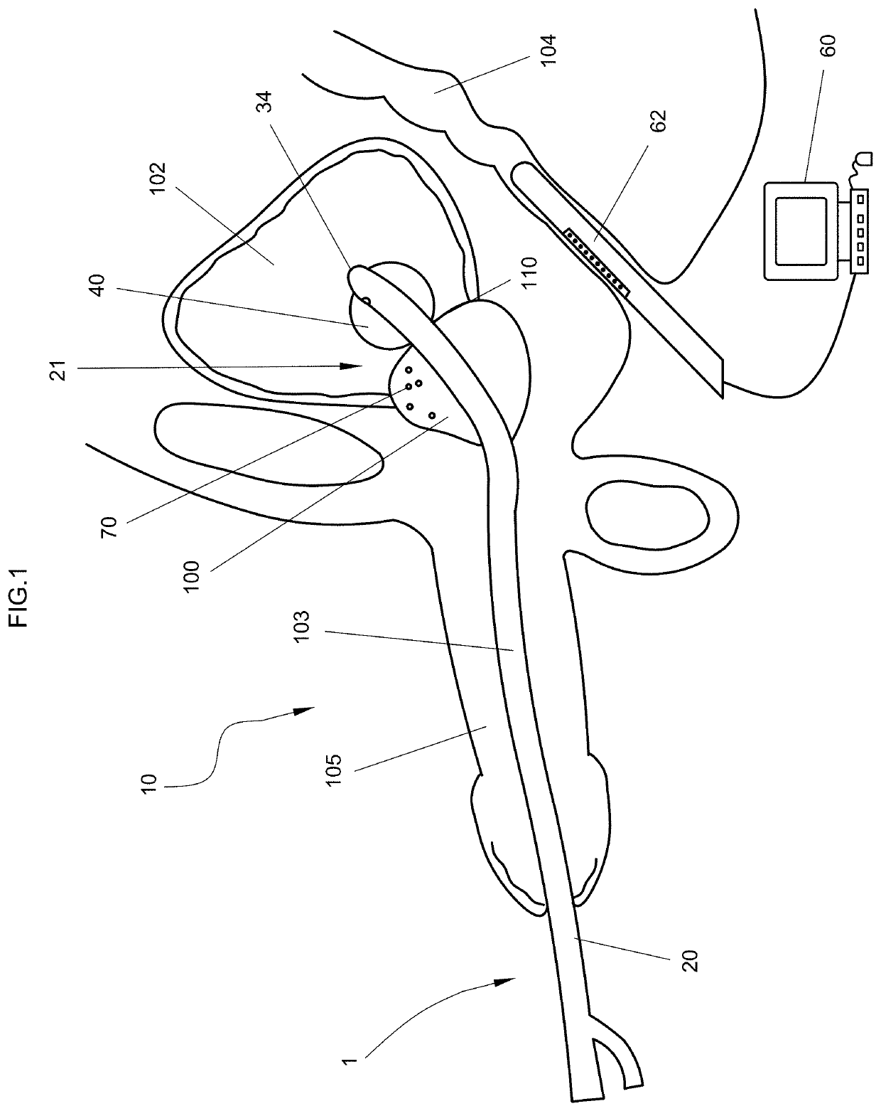
[0040]As shown in FIG. 1, the present invention is directed towards an echo-opaque identification device or a urethra identification system, generally indicated as 10, and further to a method of identifying a patient's urethral anatomical course, in real time in order to aid in the precise placement of a treatment element into the patient's prostate 100. However, the present invention is not limited to this application and can be utilized to identify any other tubular anatomic structure. A desired method of the present invention relates to the effective placement of a brachytherapy radioactive seed and / or a cryoablation probe, with precision, at a desired location within the patient's prostate 100.

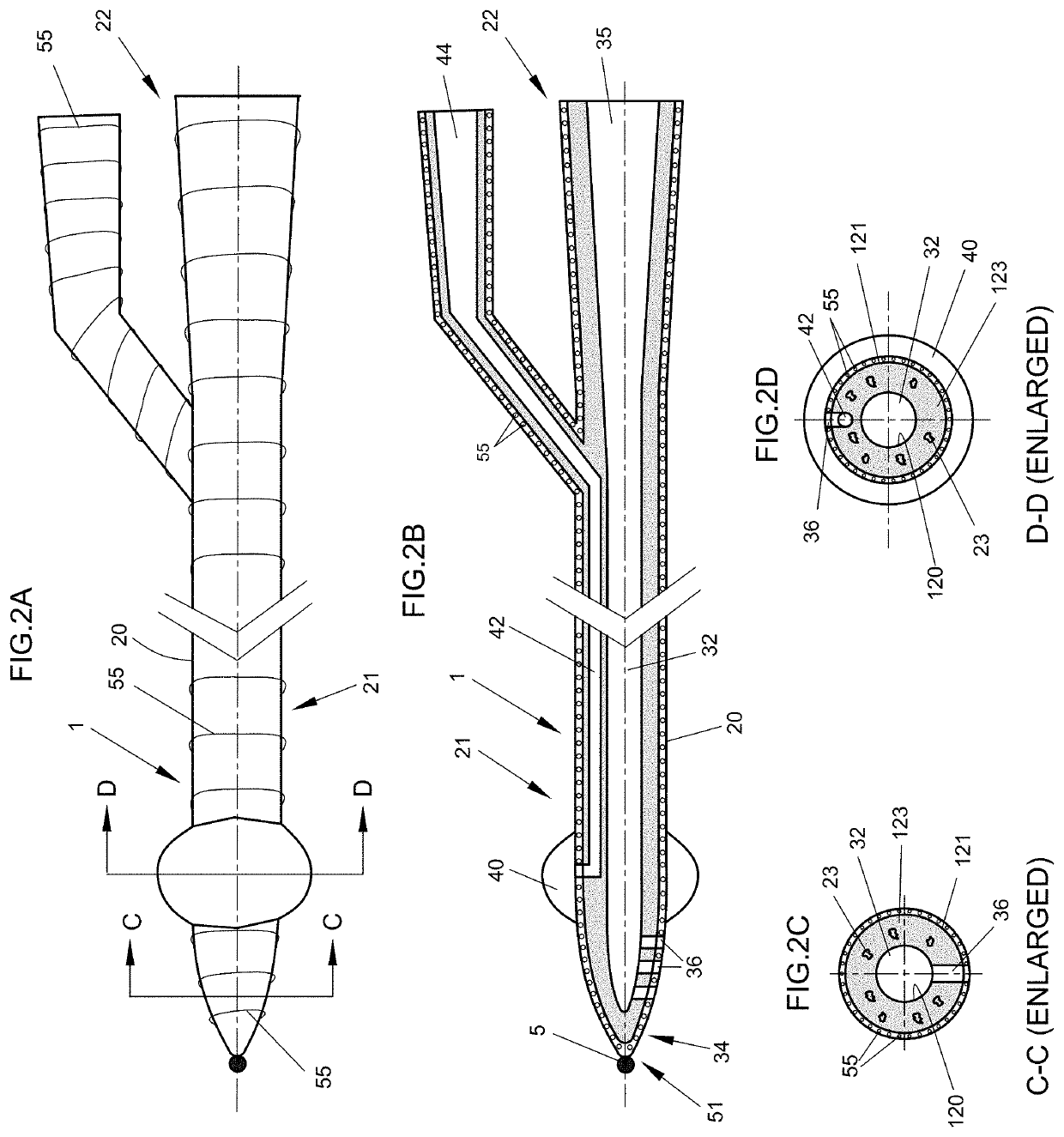
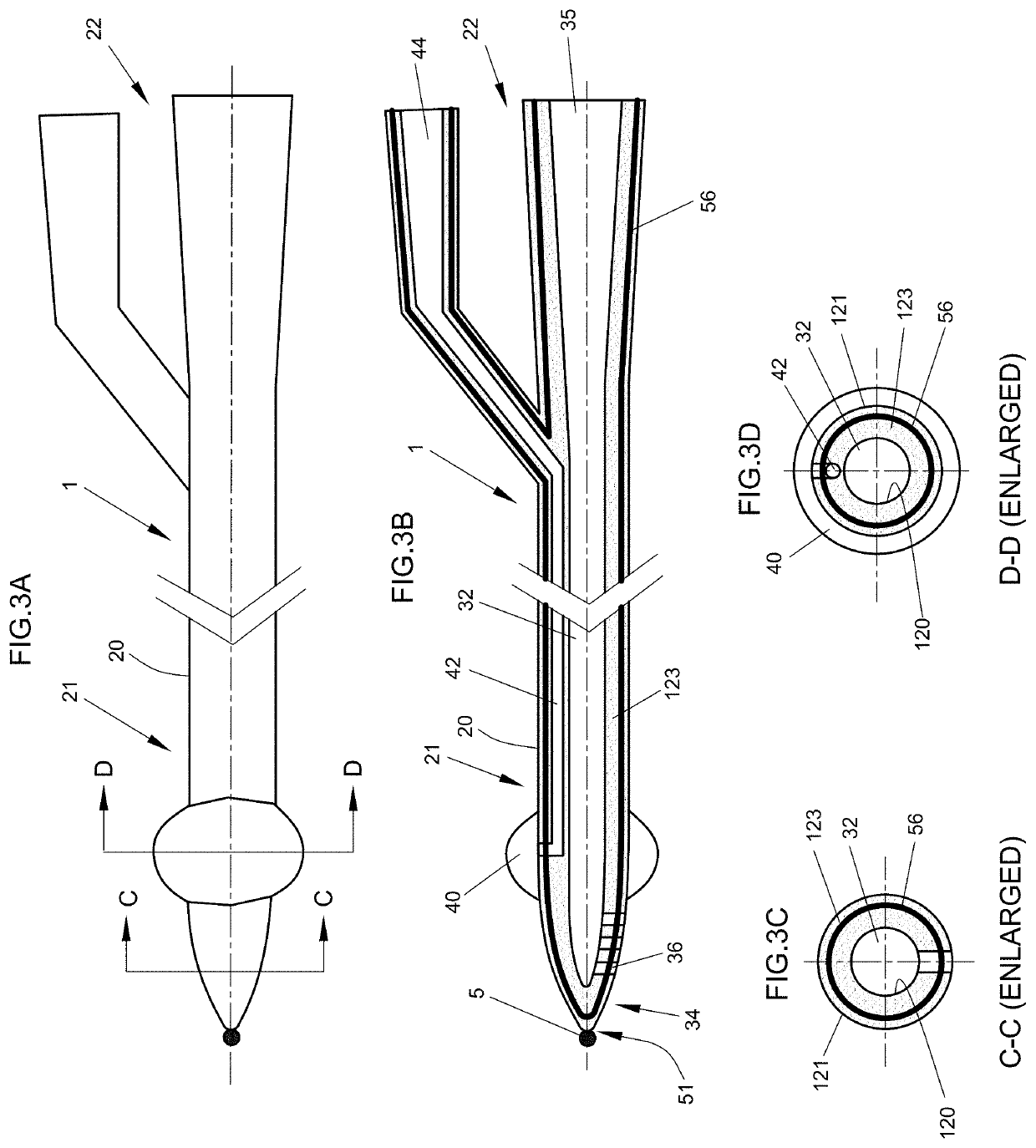
[0041]Looking first to the illustrated embodiment of the urethra identification system 10, the identification system 10 includes an echo-opaque catheter 1, such as a urethral catheter 1, and an imaging device 60. In particular, the echo-opaque catheter 1 includes an elongated flexible cath...


PUM

PropertyMeasurementUnit
flexibleaaaaaaaaaa
ultrasound imagingaaaaaaaaaa
ultrasoundaaaaaaaaaa

Abstract

A urethral catheter and its method of use to identify a patient's urethral anatomic course for the precise placement of a treatment element into the patient's prostate are provided. An echo-opaque urethral catheter including an echogenic material and a radio-opaque material in at least an image portion thereof is introduced into a urethra until its echogenic and radio-opaque image portion is generally aligned with a treatment site of the prostate. An image probe positioned relative to the treatment site is activated to obtain a real time image of the prostate and urethra by ultrasound or X-ray imaging. The echo-opaque urethral catheter includes an elongated flexible catheter, at least the image portion of which, is coated with, incorporates within or is enclosed by the echogenic and radio-opaque materials that enhance ultrasound and radio visualization.

Description

CROSS-REFERENCE[0001]This application claims priority to Provisional Application No. 60 / 807,296, filed Jul. 13, 2006, the entire content of which is hereby incorporated by reference.BACKGROUND[0002]The present invention relates to identifying a patient's anatomic urethra course and treatment devices. The invention is especially suited for accurately locating a brachytherapy or cryoablation treatment element inside a patient's prostate thereby allowing a practitioner to achieve optimal spacing between the treatment element and the urethra, while still positioning the treatment element in effective and operative proximity to a tumor, Benign Prostatic Hyperplasia (BPH) tissue, or other desired treatment site within the prostate. In this regard, the present invention is further directed towards a urethra identification system and more specifically an echo-opaque catheter, which substantially enhances the identification of the urethra at the prostate by generating, acoustic or visual int...

Claims


Application Information

Patent Timeline
Patent Type & Authority Patents(United States)
IPC IPC(8): A61B6/00A61M25/01A61B8/08A61L29/18A61L29/02A61M25/00A61B8/12A61B6/12A61M25/10A61B8/00A61F2/958
CPCA61L29/02A61M25/0108A61L29/18A61B8/0841A61B8/12A61B6/12A61M2210/166A61M25/10A61B8/445A61M25/0043
Inventor LINARES, LUISSUTHANTHIRAN, KRISHNANSUBRAMANIAN, MANNY R.
Owner BEST MEDICAL INT
Why Eureka
Try Eureka