

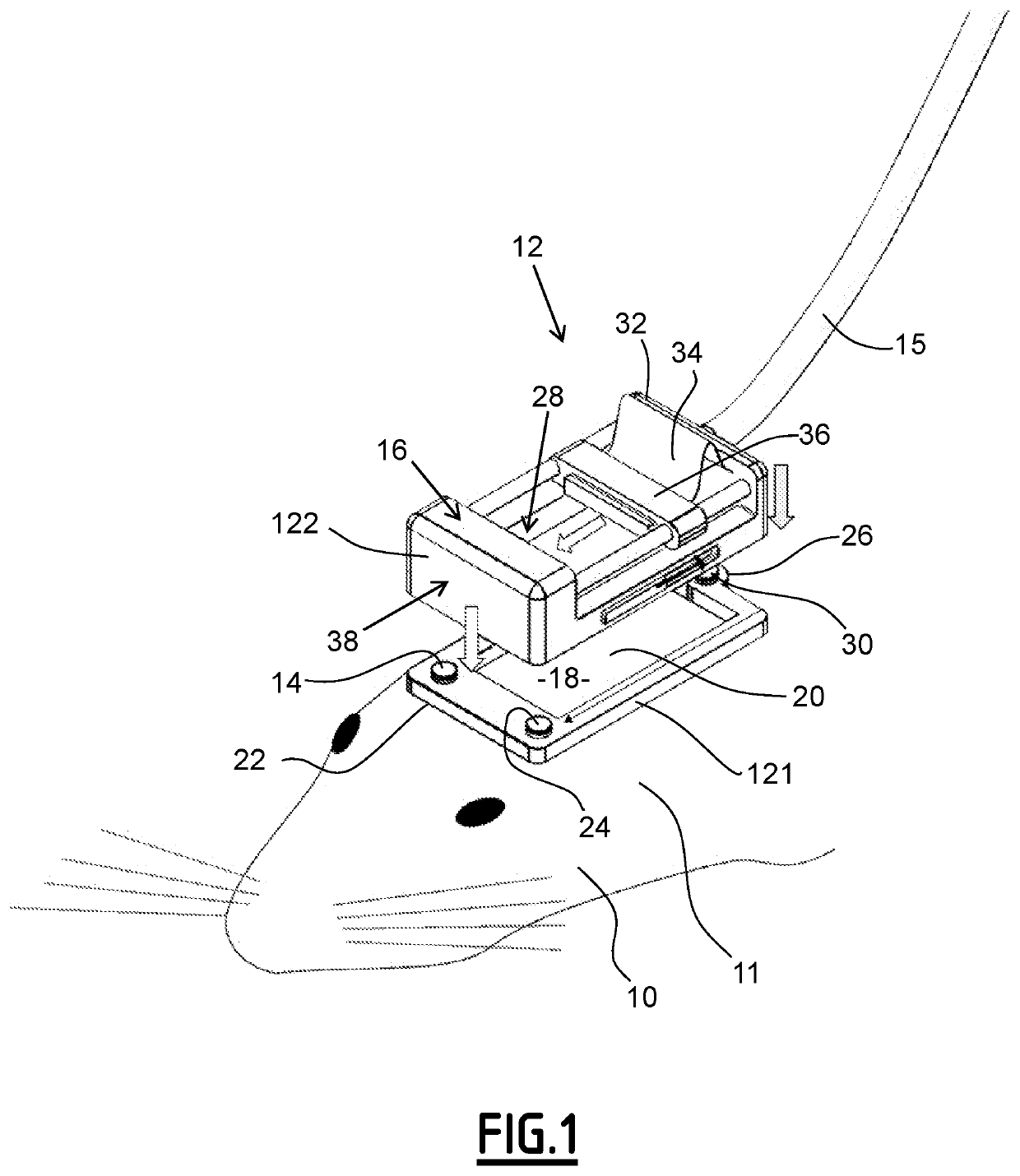
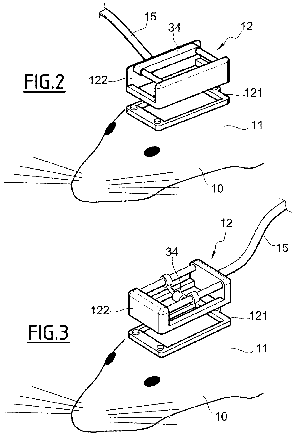
Detecting apparatus and associated imaging method
a technology of detecting apparatus and associated imaging, which is applied in the field of detecting apparatus, can solve the problems of limited sampling and inability to guaran
- Summary
- Abstract
- Description
- Claims
- Application Information
AI Technical Summary
Benefits of technology
Problems solved by technology
Method used
Image

Examples
first experiment
[0196
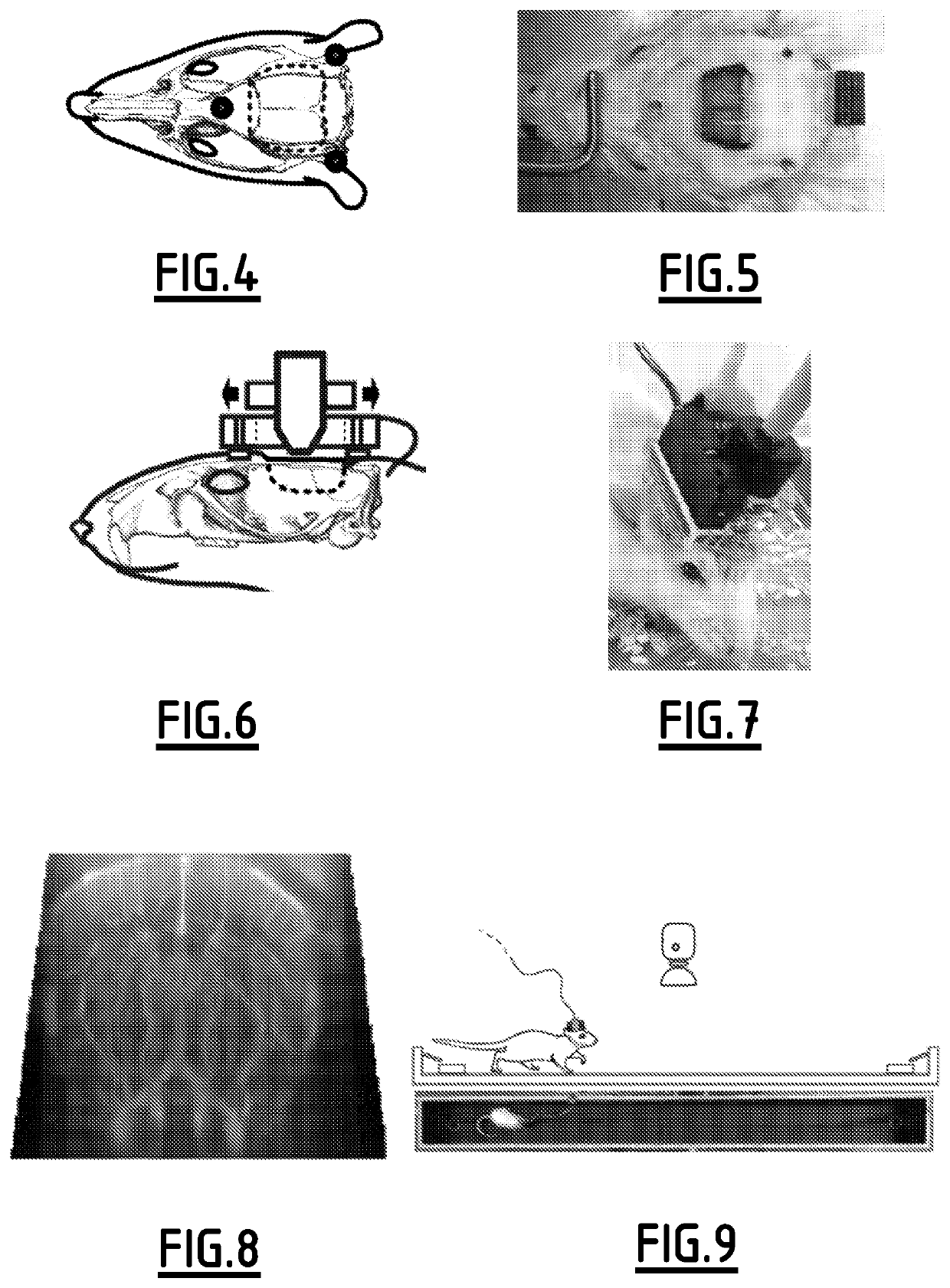
[0197]We first recorded from rats walking along a linear maze, to address how brain-wide networks activate during periods of hippocampal theta rhythm, which is a major mechanism proposed for intracerebral cross-area processing in episodic memory and spatial navigation tasks. Healthy Sprague Dawley rats (n=8) ran on a 2.25 m long, 0.2 m wide linear track for water reward. A single imaging plane included dorsal hippocampus, cortex with somatosensory areas, and thalamus. In order to temporally resolve hemodynamics as the animal crossed the maze we used a “burst mode” ultrasound sequence (see FIGS. 9 to 11), acquiring fUS compound frames at 500 Hz for 12 s. Acquisition was triggered when the animal turned around, and was followed by a 40 s lapse to collect the data. As expected, hippocampal theta was consistently associated with locomotion. Distance travelled over time was slower (56% to 64%) than in control untethered, surgery-free, rats (p−6, see FIGS. 21 to 24, tables 1 and 2). ...
second experiment
[0199
[0200]In a second experiment we scanned through the brain of an epileptic rat, to address the heterogeneous alterations in neuro-metabolic coupling during hypersynchronous seizure activity. Spontaneous generalized absence seizures were recorded from bilateral cortical electrodes in Genetic Absence Epilepsy Rats from Strasbourg (GAERS, n=12). We quantified both the relative time spent seizing and seizure duration, and found no significant difference between EEG only and EEG-mfUS conditions (see FIGS. 16 and 17 and table 3). A “continuous mode” of ultrasound acquisition was used (see FIG. 16), alternating 200 ms to generate one compound mfUS image followed with 2.8 s of processing. Multiple imaging planes were scanned for 10 min to 15 min each.
[0201]In this experiment, we also found individual pixel changes in the range from −30% to +60% (see FIGS. 12 and 13), while averaging over seizure-associated areas revealed changes from −10% to +20% (see FIGS. 18 and 19). We found distinct...
PUM
 Abstract
Description
Claims
Application Information