Implant coating composition and method of use
a technology of coating composition and coating layer, which is applied in the field of coating composition, can solve the problems of increasing the risk of adverse reactions, reducing the effect of implantation, and reducing the effect of adverse reactions
- Summary
- Abstract
- Description
- Claims
- Application Information
AI Technical Summary
Benefits of technology
Problems solved by technology
Method used
Image
Examples
example 1
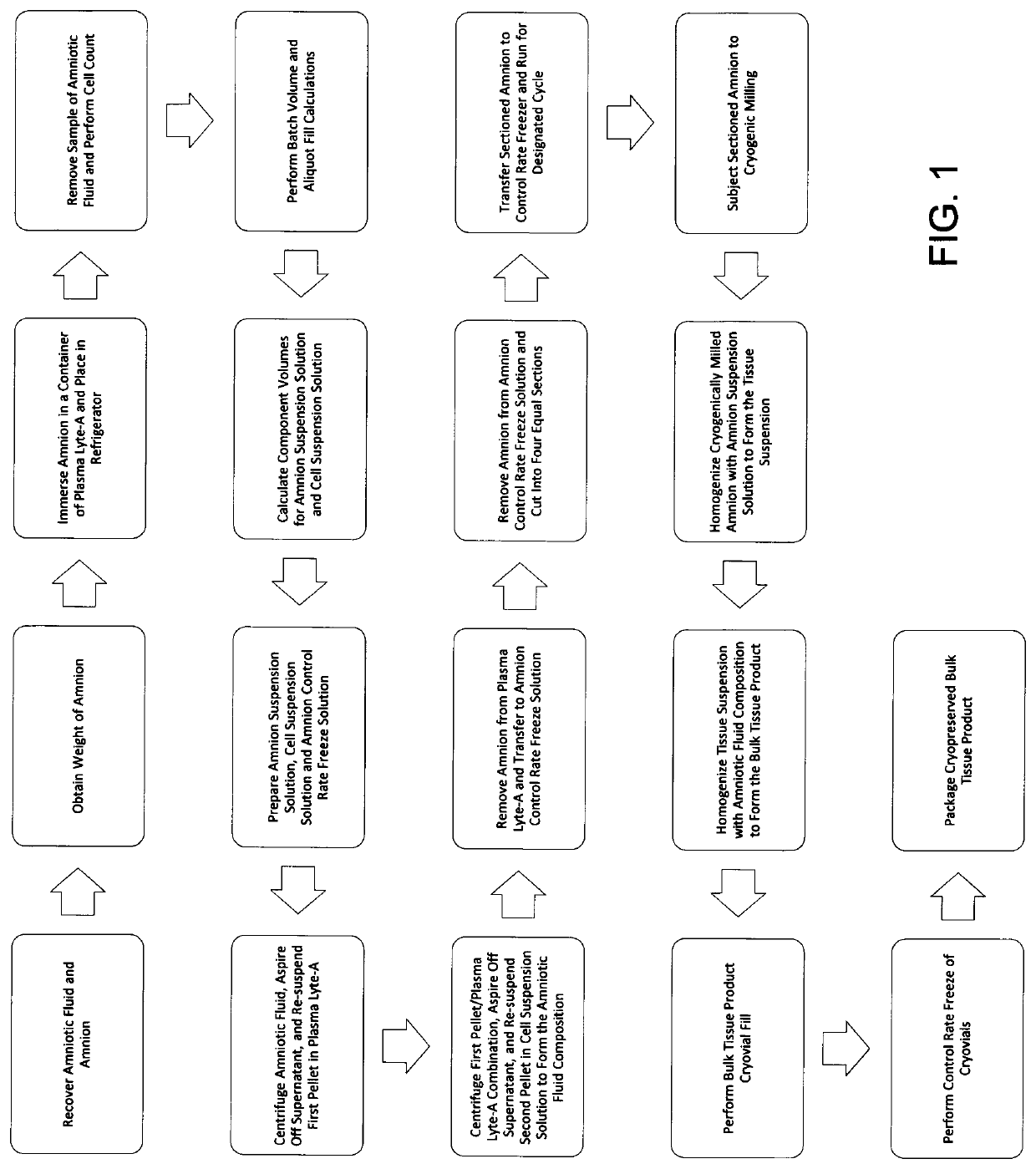
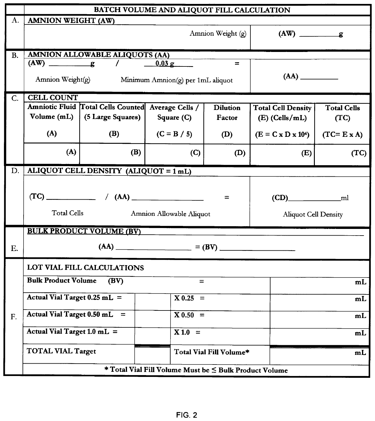
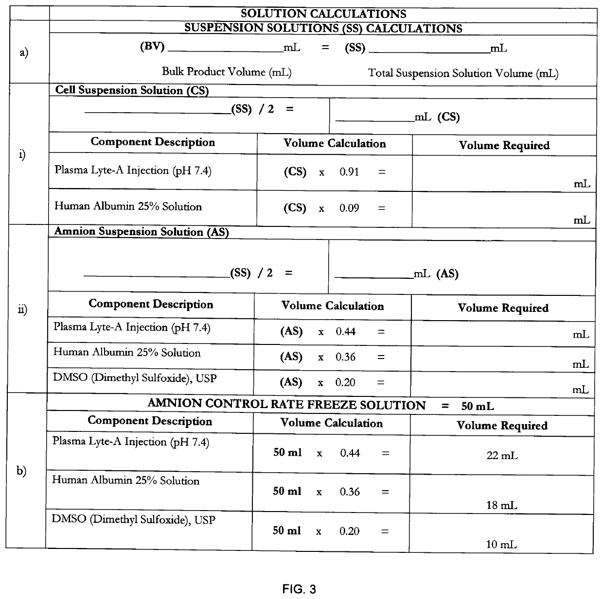
[0070]The birth tissue material coating composition may be prepared according to the method of FIG. 1, the details of which are herein provided.
[0071]Human birth tissue was obtained from a seronegative, healthy mother via Cesarean section. To maximize the overall quality of the donated tissue, a recovery technician was present in the operating room during the donor's Cesarean section to assist the surgical team with recovery, treatment and handling of the birth tissue. The donor was surgically prepped and draped per AORN standards prior to the Cesarean section procedure. The recovery technician prepared the recovery site by establishing a sterile field on a back table in the operating room.
[0072]Amniotic fluid was recovered according to the following procedures provided herein. The physician's assistant cleared all maternal blood from the surgical site. A suction cannula was positioned directly above the intended amnion / chorion membrane incision site. Using the smallest appropriate ...
example 2
[0128]A sample of a birth tissue material coating composition as prepared according to Example 1 was analyzed to assess adhesion properties to silicone breast implant materials. Sections of a silicone breast implant were cut into 10 mm square sections for all tests, with the exterior of the implant exposed to the birth tissue material coating composition, where applicable.
[0129]Phase contrast images were taken using an inverted Zeiss AxioImager fluorescence microscope coupled with an AxioCam HRm digital camera operated by AxioVision software. Silicone samples were prepared as follows: (i) incubated in the birth tissue material coating composition for five (5) minutes, then rinsed in phosphate buffered saline (PBS); (ii) coated in the birth tissue material coating composition and allowed to air dry for one (1) hour, then rinsed in PBS; or (iii) untreated silicone control. All samples were viewed through a phase contrast microscope. Observations showed subtle changes in the surface st...
example 3
[0135]To further define the in vivo effects of the birth tissue material coating composition, a model (acellular) tissue engineered scaffold derived from a decellularized human umbilical vein (dHUV) was incubated in a birth tissue material coating composition prepared in substantially the same manner as described in Example 1 and then implanted into a rat model. A control dHUV model was incubated in phosphate buffered saline (PBS), and also implanted into a rat model. After five (5) days of implantation, the scaffolds were removed and analyzed to determine effects on tissue remodeling, cell migration, fibrous capsule formation, and vascularization.
[0136]HUVs were dissected using an automated method as described in Daniel, J., Abe, K. & McFetridge, P. S. Development of the human umbilical vein scaffold for cardiovascular tissue engineering applications. ASAIO J 51, 252-261 (2005). Dissected HUV samples were decellularized in a 1% SDS (Thermo Scientific, Rockford, Ill.) solution with ...
PUM
| Property | Measurement | Unit |
|---|---|---|
| Fraction | aaaaa | aaaaa |
| Fraction | aaaaa | aaaaa |
| Fraction | aaaaa | aaaaa |
Abstract
Description
Claims
Application Information
 - R&D
- Intellectual Property
- Life Sciences
- Materials
- Tech Scout
- Unparalleled Data Quality
- Higher Quality Content
- 60% Fewer Hallucinations
Browse by: Latest US Patents, China's latest patents, Technical Efficacy Thesaurus, Application Domain, Technology Topic, Popular Technical Reports.
© 2025 PatSnap. All rights reserved.Legal|Privacy policy|Modern Slavery Act Transparency Statement|Sitemap|About US| Contact US: help@patsnap.com


