

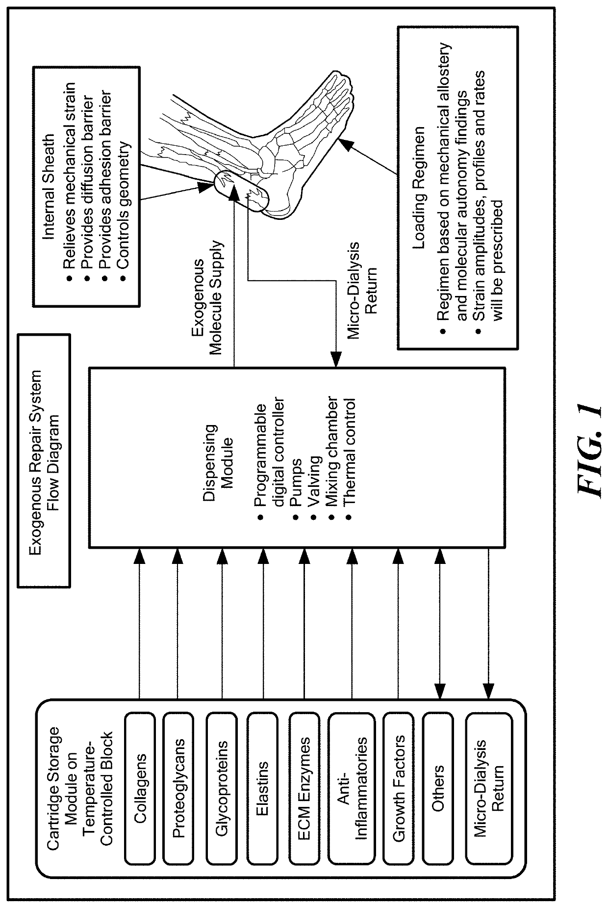
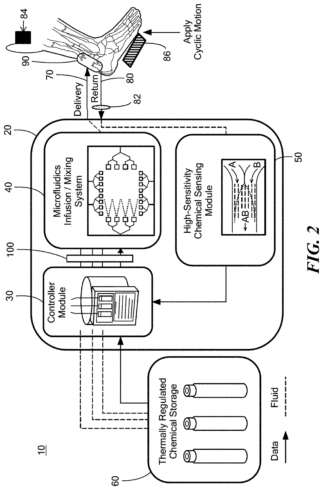
Collagenous tissue repair device
a collagenous tissue and repair device technology, applied in the field of repair of damaged biological tissues, can solve the problems of reduced molecular organization, reduced mechanical properties, inferior mobility, etc., and achieve the effect of improving the extent and quality of the restoration of damaged tissues and accelerating the repair ra
- Summary
- Abstract
- Description
- Claims
- Application Information
AI Technical Summary
Benefits of technology
Problems solved by technology
Method used
Image

Examples
example 1
Collagen Fiber Assembly by Induction of Extrinsic Strain
Collagen Preparation
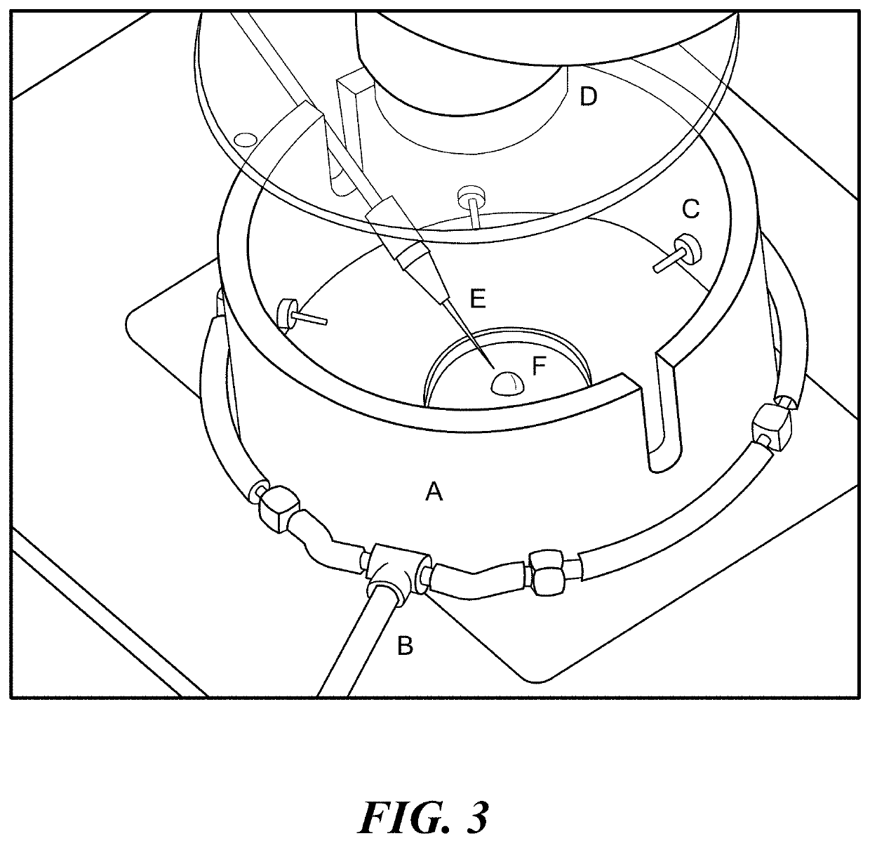
[0067]Two bovine, dermal collagen sources were used in the creation of aligned collagen fibers. Pepsin extracted collagen (5010-D, Advanced Biomatrix) and acetic acid extracted collagen (5026-D, Advanced Biomatrix) were used with a starting concentration of 6 mg / ml suspended in 0.01 M HCL. The pepsin extracted collagen was neutralized using an 8:1:1 ratio of collagen, 10× phosphate buffered saline (PBS) (BP399-1, Fisher Scientific), and 0.1 M NaOH (12419-0010, Fisher Scientific) respectively. This resulted in a pH of 7.3. For the experiments using decorin (D8428-.5MG, Sigma Aldrich), a ratio of 2% decorin to collagen molar ratio was used. The 0.5 mg lyophilized decorin was first reconstituted with 1 ml deionized water. To prevent any spontaneous polymerization while working with acetic acid extracted collagen, the neutralization ratio was altered such that the amount of 0.1 M NaOH was increased to 115%. Cons...
PUM
| Property | Measurement | Unit |
|---|---|---|
| concentration | aaaaa | aaaaa |
| diameter | aaaaa | aaaaa |
| concentration | aaaaa | aaaaa |
Abstract
Description
Claims
Application Information