

Use of engineered renal tissues in assays
a technology of renal tubules and engineered tissues, applied in the field of renal tubule models, can solve the problems of inability to accurately predict organ-specific toxicity, and damage to the tubular epithelium and surrounding cells, so as to facilitate the correct localization of drug transporters, facilitate optimal morphology and function, and accurately study
- Summary
- Abstract
- Description
- Claims
- Application Information
AI Technical Summary
Benefits of technology
Problems solved by technology
Method used
Image

Examples
example 1
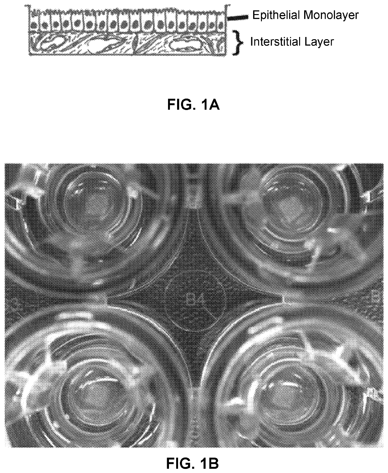
A Bioprinted Three-Dimensional Renal Tubule Cell Model
[0416]Human umbilical vein endothelial cells (HUVEC) were purchased from BD
[0417]Biosciences (Franklin Lakes, N.J.) and cultured in EGM-2 media with EBM-2 supplements without gentamycin or amphotericin B (Lonza, Basel, Switzerland). Adult renal fibroblasts were purchased from DV Biologics (Yorba Linda, Calif.) and grown in Fibroblast Cellutions Medium with Fibroblast Cellutions supplement (DV Biologics, Yorba Linda, Calif.). Primary human RPTEC were purchased from four different commercial vendors (Lonza, Sciencell (Carlsbad, Calif.), Zen-Bio (Research Triangle Park, N.C.), Lifeline Cell Technology (Frederick, Md.) and cultured according to the manufacturer's instructions.
[0418]All kidneys were ethically sourced through the National Disease Research Interchange (Philadelphia, Pa.). RPTEC cells were isolated as previously described (Vesey et al., 2009). In brief, upon receipt, kidneys were aseptically unpacked and cleaned to remov...
example 2
A Bioprinted Three-Dimensional Renal Tubule Cell Model
[0419]3D PT Novo View™ Tissues were fabricated as described (Nguyen et al., 2016). Briefly, cultured renal fibroblasts and HUVEC were combined in a 50:50 ratio and resuspended in NovoGel® Bio-Ink, and then bioprinted onto 0.4 μm Transwell® clear polyester membrane inserts in a 24-well plate (Coming Costar, Coming, NY) using a NovoGen Bioprinter® Instrument (Organovo Inc., San Diego, Calif.) with previously established protocols (Forgacs et al., 2012; Murphy et al., 2015; Nguyen et al., 2016). Following bioprinting, NovoView′ Tissues were cultured in NovoView™ Kidney Media (Organovo, San Diego, Calif.). On culture day 3, primary RPTEC cells were added to the tissues in a suspension of 1.25×10⁶ cells / ml in RPTEC media. Tissues were then maintained for up to 30 days in Novo View™ Kidney Media (Organovo, San Diego, Calif.), with media exchanges every other day. For toxicity studies, tissues were dosed daily to the apical and basolate...
example 3
Metabolic and Viability Assays on Bioprinted Tissues
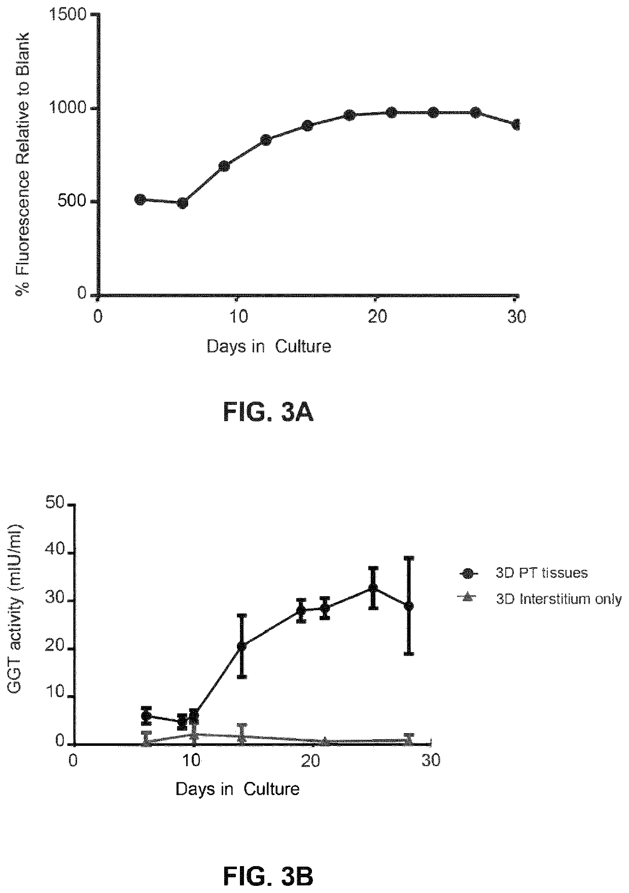
[0420]Assessment of metabolic activity as a surrogate for tissue viability and health was performed by alamarBlue™ assay according to the manufacturer's protocol (Thermo Fisher, Carlsbad, Calif.). Briefly, tissues were washed twice with Dulbecco's phosphate buffered saline (DPBS), and RPTEC media supplemented with 10% v / v alamarBlue′ reagent was added to each tissue. All tissues were incubated for 2 hours at 37° C. with 95% relative humidity and 5% CO2. After incubation, the alamarBlue′ solution was removed and fluorescence was measured on a BMG Labtech POLARstar® Omega reader (Cary, N.C.) with an excitation filter of 560 nm and an emission filter of 590 nm. Graphed data represent the percent relative fluorescence units (RFU) compared to blank for metabolic activity over time, or the percent RFU compared to vehicle control for toxicity studies.
[0421]Lactate dehydrogenase (LDH) activity assay was performed according to the manufactu...
PUM
| Property | Measurement | Unit |
|---|---|---|
| thick | aaaaa | aaaaa |
| thick | aaaaa | aaaaa |
| thick | aaaaa | aaaaa |
Abstract
Description
Claims
Application Information