Engineered renal tissues, arrays thereof, and methods of making the same

Active Publication Date: 2016-04-07
ORGANOVO
View PDF1 Cites 6 Cited by
  • Summary
  • Abstract
  • Description
  • Claims
  • Application Information

AI Technical Summary

Benefits of technology

[0004]The engineered tissues described herein represent a model of the tubulointerstitial interface in which human renal interstitial tissue is supporting human renal proximal tubule epithelial cells to facilitate their optimal morphology and function. Creation of a three-dimensional tubulointerstitial interface facilitates the correct localization of drug transporters and receptors required for metabolism in order to accurately study how small molecule, chemicals, contaminants, or biologics affect the renal proximal tubule. This represents a more physiologically relevant alternative to two-

Problems solved by technology

Current in vitro renal toxicity models, such as monolayers of epithelial cells and in vivo models, such as live rodents, cannot effectively predict a drug's effects, toxicity, or metabolism in humans.
For example, conventional renal cell culture models lack the complexity of native

Method used


Image

  • Engineered renal tissues, arrays thereof, and methods of making the same
  • Engineered renal tissues, arrays thereof, and methods of making the same
  • Engineered renal tissues, arrays thereof, and methods of making the same

Examples


example 1

A Bioprinted Three-Dimensional Renal Tubule Cell Model

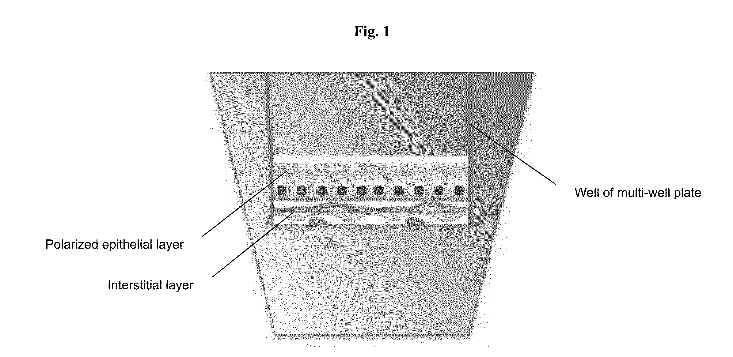
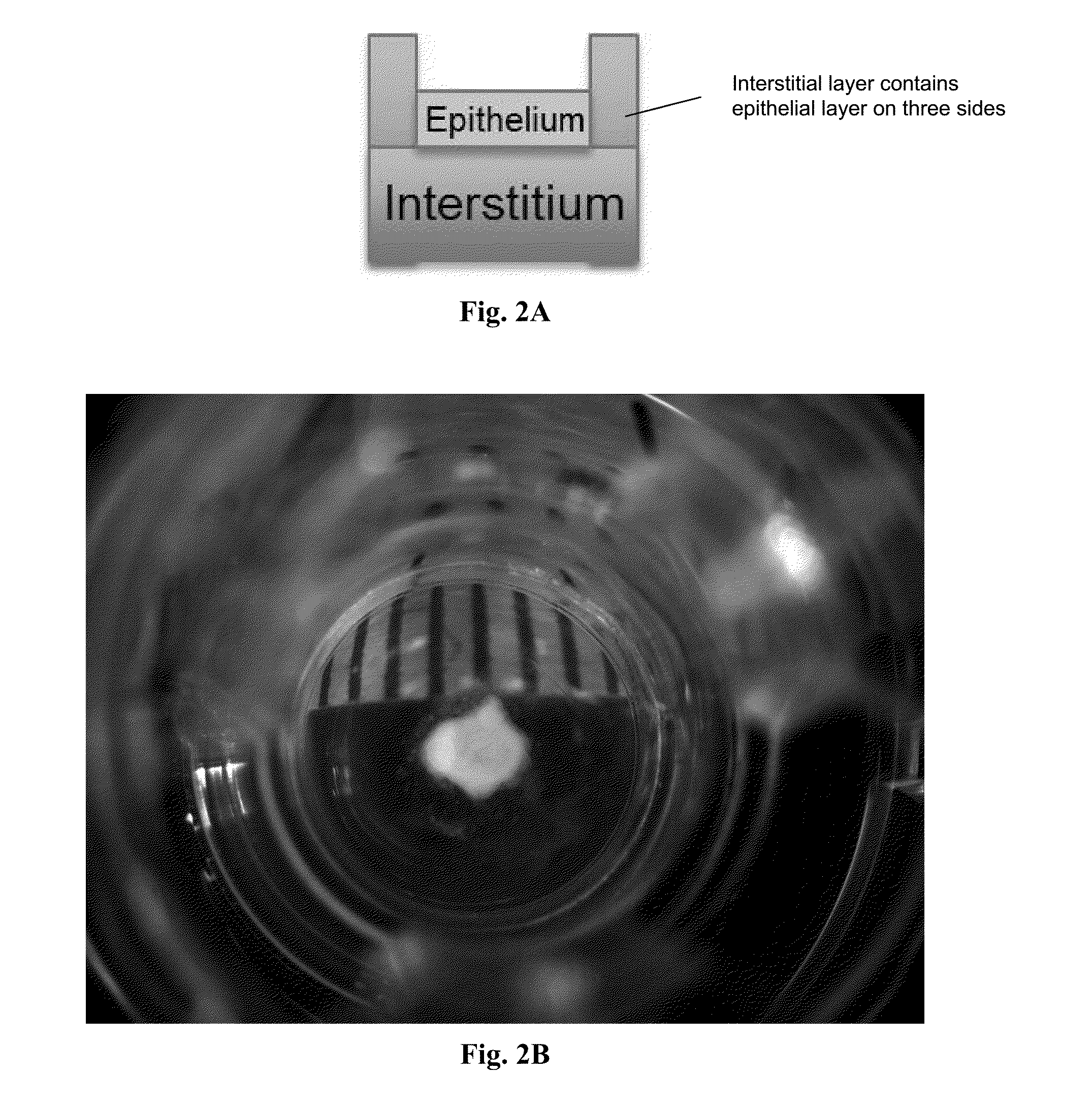
[0099]A 3-layer cup shape with dimensions 3 mm×3 mm×0.75 mm was bioprinted onto a Transwell membrane in a 12-well tissue culture plate (FIGS. 2A and 2B). The bottom sheet and two layered walls were composed of 75% adult renal fibroblasts (aRF) and 25% human umbilical vein endothelial cells (HUVEC). The cells were resuspended at 150 million cells / ml in Novogel® 2.0. The cup was filled with a dilute suspension of 100% epithelial cells (MDCK) at 1-5 million cells / ml in 2% gelatin (“Structure 1”). Immediately following bioprinting, structures were surrounded with molten Novogel® 1.0 and were then cultured in a mixture of renal fibroblasts media, HUVEC media, and MDCK media.

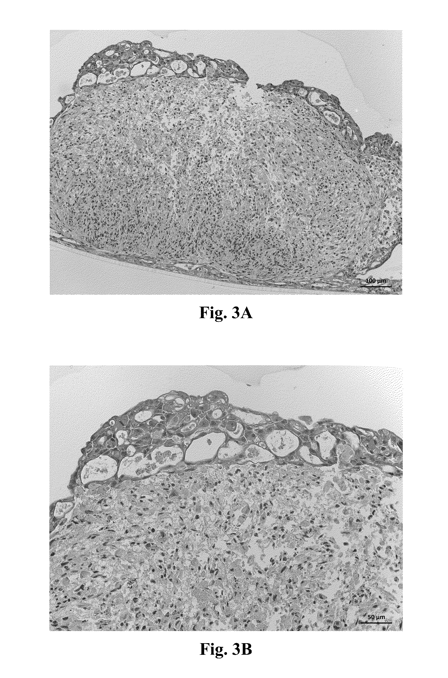
[0100]Assessment of Structure 1 was performed by histological staining for cell-type specific markers. A representative H&E stain for Structure 1 is shown in FIGS. 3A and 3B. Constructs were stained for CD31 and TE7 to assess the relative positions of the HUVEC cel...

example 2

A Bioprinted Three-Dimensional Renal Tubule Cell Model

[0101]A sheet with dimensions 3 mm×3 mm×250 μm was bioprinted onto a Transwell membrane in a 12-well plate. The tissue sheet was composed of 75% adult renal fibroblasts and 25% HUVEC resuspended at 150 million cells / ml in Novogel® 2.0. The edges of the sheet were bordered by a 500 μm thick bioprinted hydrogel-only wall composed of Novogel® 3.0. Immediately following bioprinting, the boundary wall was crosslinked with 50 mM calcium chloride for 2 minutes. This solution was then aspirated and the constructs were surrounded with molten Novogel® 1.0. A dilute suspension of epithelial cells (MDCK or hTERT-RPTEC) at 1 million cells / ml was then added to the top surface of the structure by deposition using an inkjet spray module set with a 100 ms valve open time (“Structure 2”). A schematic of Structure 2 is shown in FIGS. 6A and 6B. Use of the inkjet spray module allows for deposition of epithelial cells immediately after printing or in...

example 3

A Bioprinted Three-Dimensional Renal Tubule Cell Model

[0103]To reduce the thickness and cellularity of the interstitial layer, the cell ratio was changed to 50% fibroblasts / 50% HUVEC the concentration of the cells was 1.25×10⁸ cells / mL. The interstitial layer of the renal proximal tubule model is composed of renal fibroblasts and HUVECs in Novogel® 2.0. Constructs produced in this example show proximal tubule epithelial cells (RPTECs) in 3D renal tissues that exhibit features of polarization. E-cadherin (green) at the lateral membranes between RPTECs corresponds to tight junctions (FIGS. 9A, B and C). Further, the basement membrane corresponding to the interstitial layer produces collagen (FIGS. 9 A and B; stained with red). H & E staining is shown (FIG. 10A), and brush borders are indicated (FIG. 11A, arrows). Trichrome staining indicates collagen secretion (FIGS. 10B, blue and 11B, arrows), and CD31 staining indicates the presence of HUVEC networks (FIG. 12, asterisks). Bioprinted...


PUM


Abstract

Disclosed are renal tissues and arrays thereof that include a layer of renal interstitial tissue, the renal interstitial tissue comprising renal fibroblasts and endothelial cells; and a layer of renal epithelial tissue, the renal epithelial tissue comprising renal tubular epithelial cells, the renal epithelial tissue in contact with the layer of renal interstitial tissue to form a three-dimensional, engineered, biological renal tissue. Also disclosed are methods of fabricating and using the same.

Description

CROSS-REFERENCE TO RELATED APPLICATIONS[0001]This application claims the benefit of U.S. Patent Application No. 62 / 060,416, filed Oct. 6, 2014, and U.S. Patent Application No. 62 / 140,285, filed Mar. 30, 2015, the entire disclosures of which are hereby incorporated herein by reference.BACKGROUND OF THE INVENTION[0002]The full cost of bringing a new drug to market—from discovery through clinical trials to approval—is typically hundreds of millions or billions of U.S. dollars. This is in part because ninety-five percent of the experimental medicines that are studied in humans fail to be both effective and safe. Renal toxicity is a major cause of drug attrition at the clinical trial stage, and the primary site of this toxicity is within the proximal tubule.SUMMARY OF THE INVENTION[0003]Current in vitro renal toxicity models, such as monolayers of epithelial cells and in vivo models, such as live rodents, cannot effectively predict a drug's effects, toxicity, or metabolism in humans. For...

Claims


Application Information

Patent Timeline
IPC IPC(8): C12N5/071G01N33/50
CPCC12N5/0684G01N33/5014G01N33/5044C12N5/0697C12N2502/256C12N2502/28C12N2502/1323G01N2500/02G01N2500/10
Inventor NGUYEN, DEBORAH LYNN GREENEKING, SHELBY MARIEPRESNELL, SHARON C.
Owner ORGANOVO
Why Eureka
Try Eureka