Tissue preparation using nuclease
a technology of nucleotide and tissue, applied in the field of tissue preparation using nuclease, can solve the problems of false detection of dna mutations in cancer diagnosis, udg treatment fails to cure apparent c:g>t:a single-nucleotide change, and cancer is a major health risk in the united states and internationally, so as to improve the analysis of dna and reduce artifacts
- Summary
- Abstract
- Description
- Claims
- Application Information
AI Technical Summary
Benefits of technology
Problems solved by technology
Method used
Image
Examples
example 1
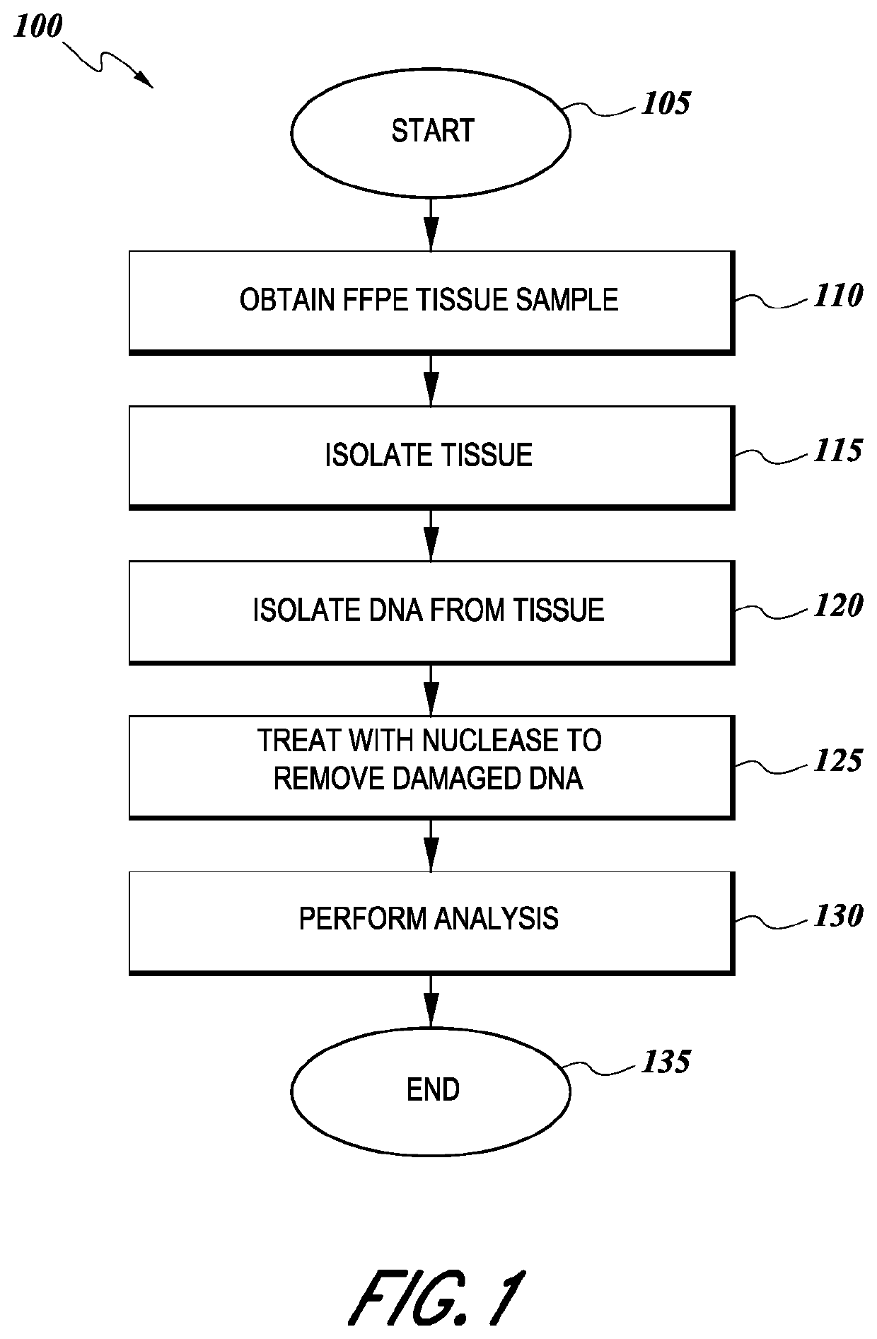
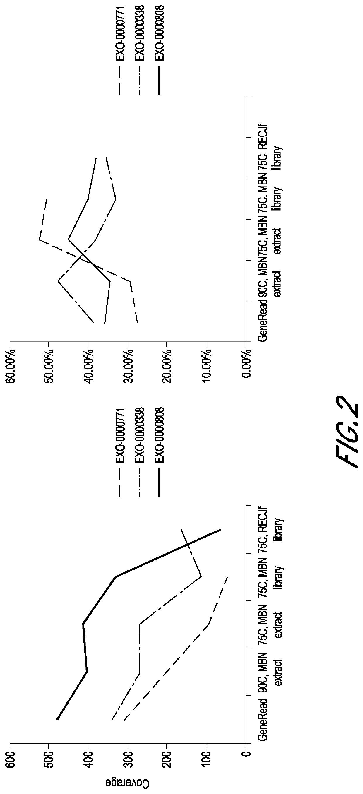
[0046]Three FFPE blocks of normal tissues of low quality (EXO-0000771), moderate quality (EXO-0000338), and high quality (EXO-0000808), respectively, were selected. Excess paraffin was trimmed off from each block. Each FFPE block was sectioned using a microtome to obtain five 4 μm slides. Each slide was placed into a separate microcentrifuge tube.
example 2
DNA Extraction and Library Preparation—Group A
[0047]Three FFPE slides of low quality, moderate quality, and high quality, respectively, were processed according to the DNA purification protocol of Qiagen GeneRead DNA FFPE Kit, followed by the library construction protocol of KAPA HyperPlus Kit.
[0048]Briefly, the slides were dewaxed by warming at 56° C. in 160 μL Deparaffinization Solution for 3 minutes. The tissues were lysed at 56° C. for 1 hour in 55 μL RNase-free water, 25 μL Buffer FTB, and 20 μL proteinase K. The tissues were then heat-treated at 90° C. for 1 hour to de-crosslink. After the heat treatment, the tissues were centrifuged and transferred to new microcentrifuge tubes. Then, 115 μL RNase-free water and 35 μL of the enzyme uracil-N-glycosylase (UNG) were added to the samples, and the samples were incubated at 50° C. for 1 hour to remove artifacts of deaminated cytosine residues of the FFPE DNA. The samples were then treated with 2 μL RNase A at room temperature for 2 ...
example 3
DNA Extraction and Library Preparation—Group B
[0051]Three FFPE slides of low quality, moderate quality, and high quality, respectively, were processed according to the DNA purification protocol of Qiagen GeneRead DNA FFPE Kit, followed by the library construction protocol of KAPA HyperPlus Kit, but with some modifications.
[0052]Briefly, the slides were dewaxed by warming at 56° C. in 160 μL Deparaffinization Solution for 3 minutes. The tissues were lysed at 56° C. for 1 hour in 55 μL RNase-free water, 25 μL Buffer FTB, and 20 μL proteinase K. The tissues were then heat-treated at 90° C. for 1 hour to de-crosslink. After the heat treatment, the tissues were centrifuged and transferred to new microcentrifuge tubes. Then, 115 μL RNase-free water and 35 μL of the enzyme uracil-N-glycosylase (UNG) were added to the samples to remove artifacts of deaminated cytosine residues of the FFPE DNA. In contrast to Group A, here the sampled were incubated with UNG at 50° C. for 30 minutes. After i...
PUM
| Property | Measurement | Unit |
|---|---|---|
| Efficiency | aaaaa | aaaaa |
| Refractory | aaaaa | aaaaa |
Abstract
Description
Claims
Application Information
 - R&D
- Intellectual Property
- Life Sciences
- Materials
- Tech Scout
- Unparalleled Data Quality
- Higher Quality Content
- 60% Fewer Hallucinations
Browse by: Latest US Patents, China's latest patents, Technical Efficacy Thesaurus, Application Domain, Technology Topic, Popular Technical Reports.
© 2025 PatSnap. All rights reserved.Legal|Privacy policy|Modern Slavery Act Transparency Statement|Sitemap|About US| Contact US: help@patsnap.com


