Heparin-binding growth factor (HBGF) polypeptides
a growth factor and polypeptide technology, applied in the field of growth factors, can solve the problem that ctgf alone cannot induce this property in fibroblasts
- Summary
- Abstract
- Description
- Claims
- Application Information
AI Technical Summary
Problems solved by technology
Method used
Image
Examples
example 2
HBGF Polypeptide Sequencing
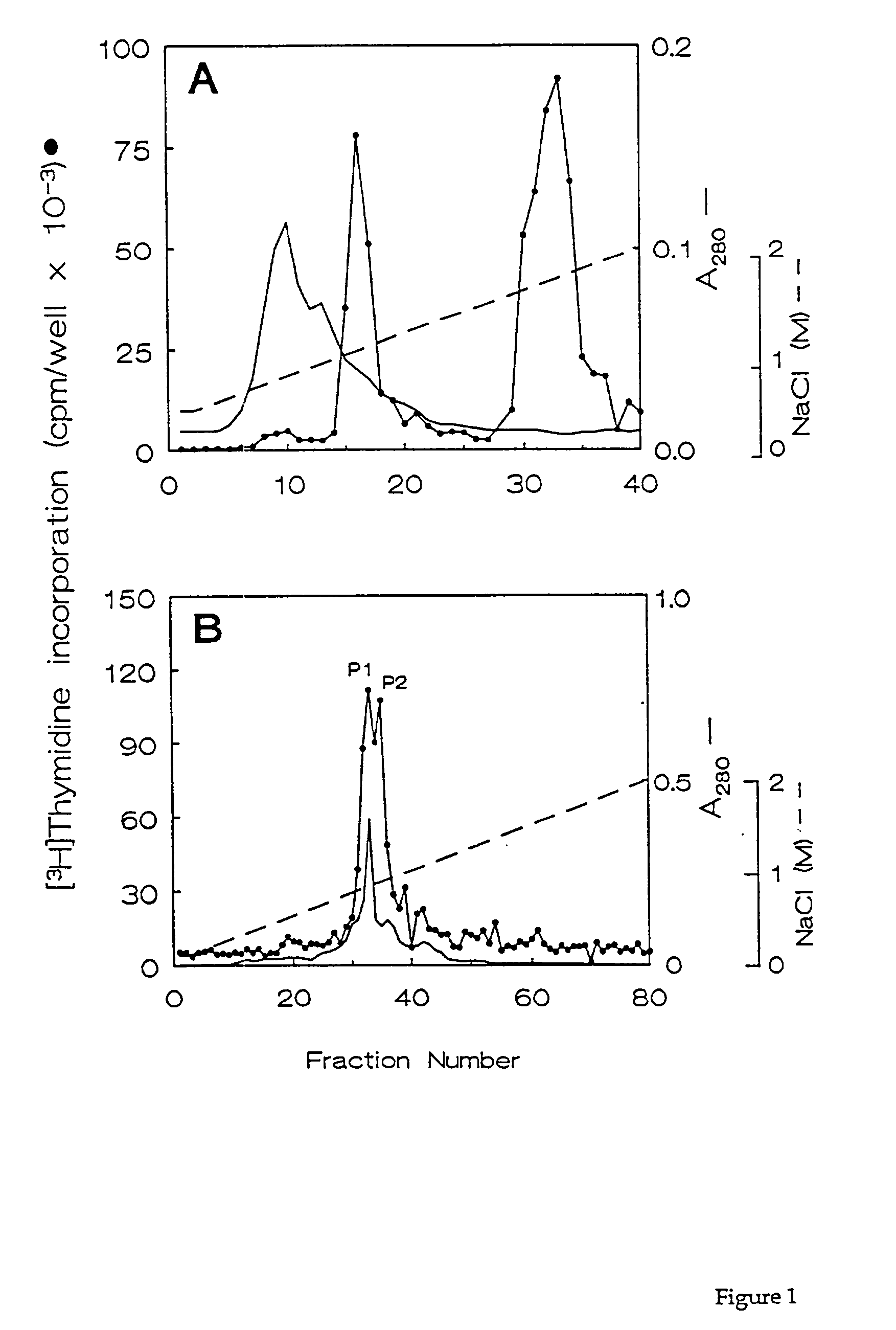
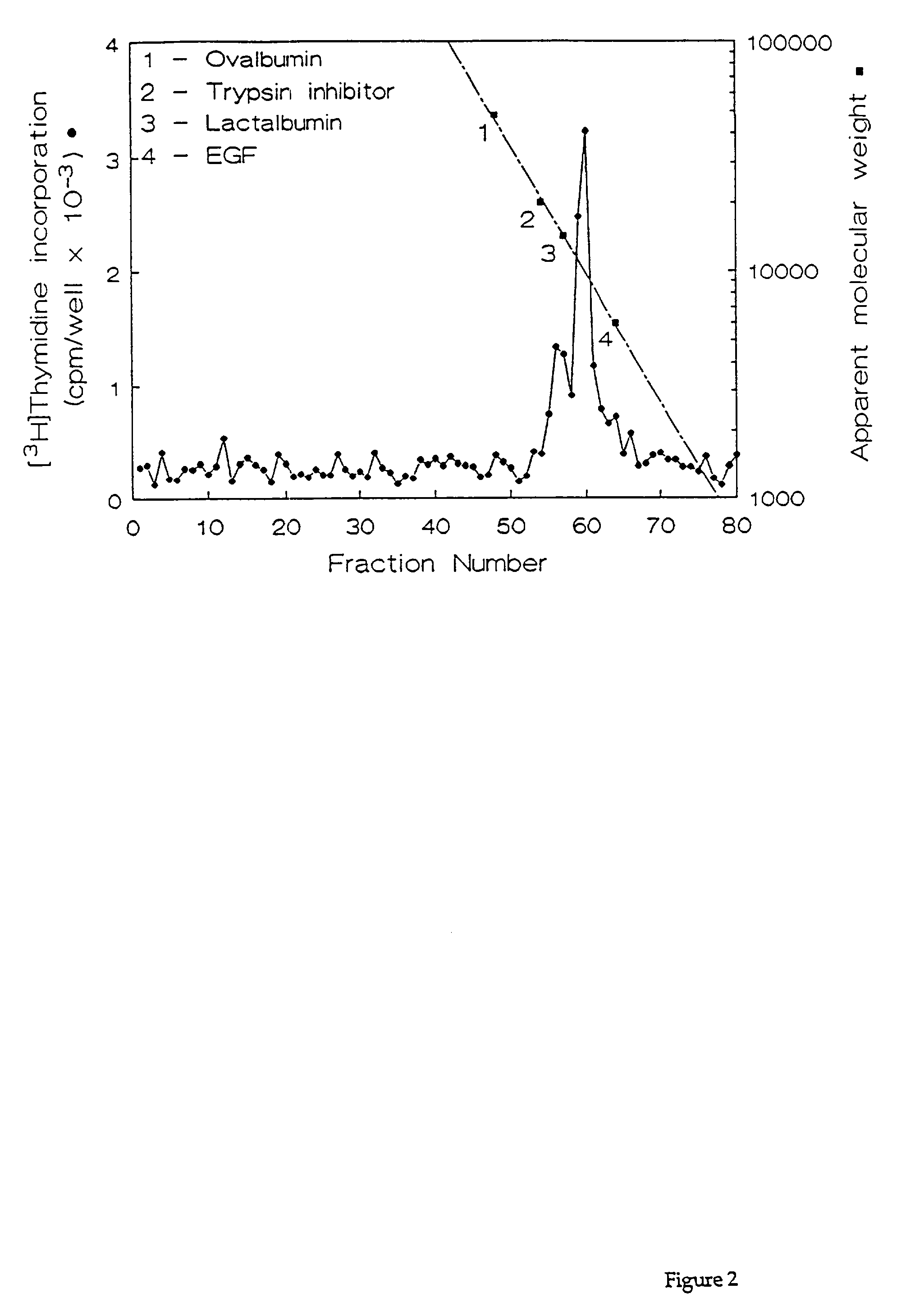
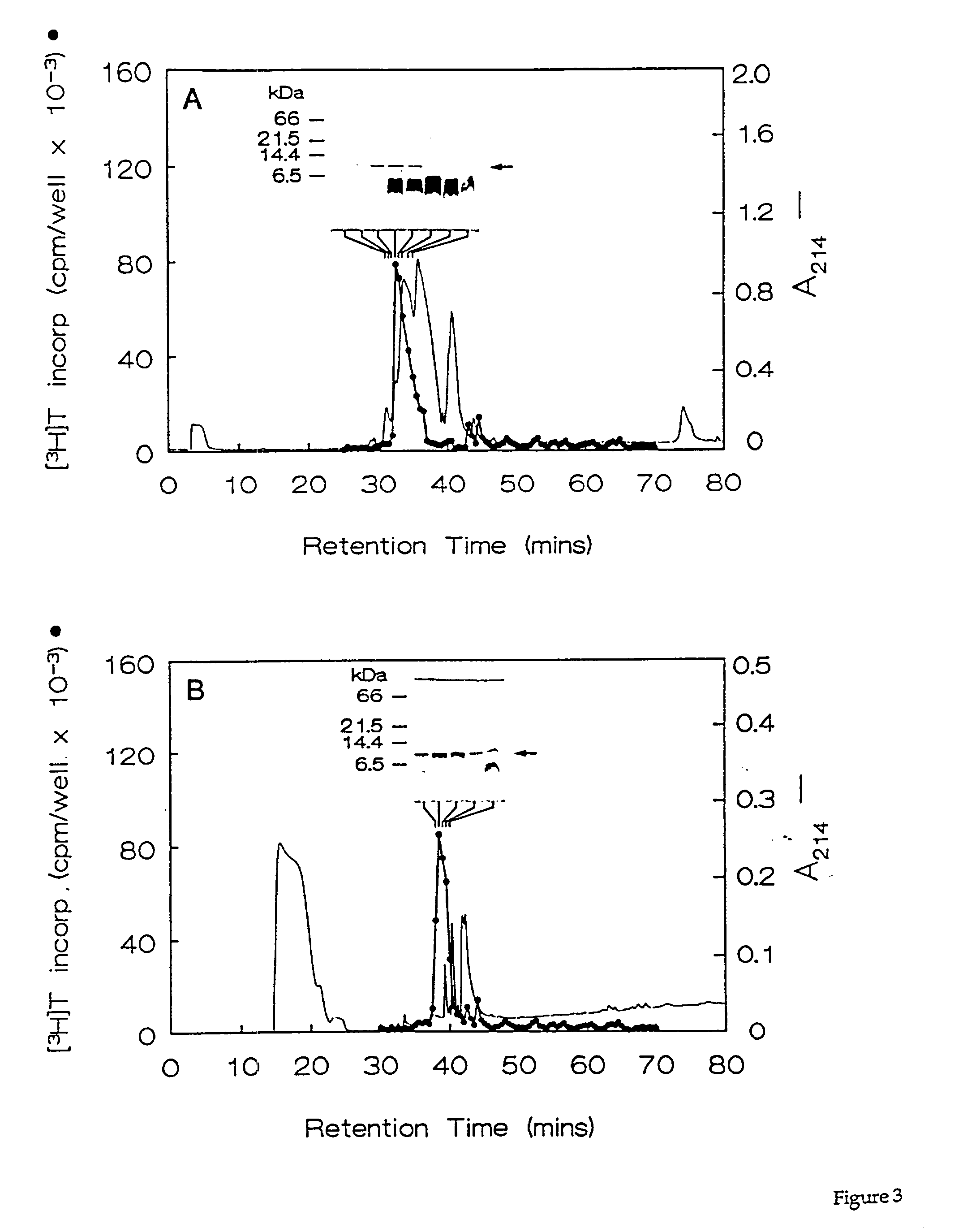
[0066] Fractions containing the HPLC purified growth factors were pooled, dried, and subjected to preparative SDS-PAGE. Proteins in the gel were transferred for 90 min. at 300 mA to a polvinylidene difluoride membrane using 10 mM CAPS buffer (pH 11). The location of the proteins of interest was determined by staining the blots with 0.1% Coomassie R250 in 50% methanol for 2 min, followed by destaining with 50% methanol, 10% acetic acid. Half of each 10-kDa protein band was excised and submitted for N-terminal amino acid sequencing on a model 470A gas phase sequenator (Applied BioSystems, Foster City, Calif.). Phenylthiohydantoin-derivative-s were identified by C.sub.18 reverse phase HPLC. A 16-residue sequence was obtained for HBGF-0.8-P1 with an undetermined residue at position 10, and a 12-residue sequence was obtained for HBGF-0.8-P2 with an undetermined residue at position 9 (Table 2). These data showed that HBGF-0.8-P1 and -P2 were N-terminally identic...
example 3
HBGF Antibody Production
[0070] Since HBGFs represent microheterogenous forms of truncated CTGF, the relationship of HBGF to CTGF was investigated. The presence of the 10-kDa protein in the starting material was confirmed by Western blotting of unfractionated ULF samples using a CTGF antibody that reacted with HPLC-purified HBGF polypeptides.
[0071] To produce the antibody, a four-branched multiple antigenic CTGF-(247-260) peptide comprising the sequence EENIKKGKKCIRTP (residues 247-260) (SEQ ID NO: 5) was produced on a Synergy 432A peptide synthesizer (Applied BioSystems) and purified by reverse-phase HPLC using a C.sub.8 column (0.46.times.36 cm; Rainin Instruments) that was developed with a 90-min 5-95% acetonitrile gradient in water, 0.1% trifluoroacetic acid. Fractions containing the purified polypeptides were pooled, evaporated to dryness, and reconstituted in sterile water. Two New Zealand White rabbits (rabbits A and B), which had been bled to collect preimmune serum, were inj...
example 4
Generation of the 10-kDa HBGF Polypeptides
[0072] Western blotting was performed as has been previously described (Harlow and Lane, 1988, Antibodies, A Laboratory Manual, Cold Spring Harbor Laboratory, New York, Current Edition). Briefly, SDS-PAGE was performed under reducing conditions using 18% polyacrylamide mini-gels as described (Kim, G. Y., et al., Biol. Reprod. 52, 561-571 (1995)). Silver staining of proteins was performed as described (Wray, W., et al., Anal. Biochem. 118, 197-203 (1981)). Western blotting was performed on (i) HPLC-purified growth factors, (ii) 8 .mu.l of unfractionated ULF, or (iii) 100 .mu.l of ULF after passage through 20-.mu.l beds of heparin-Sepharose in the presence of 10 mM Tris-HCl, 0.5 M NaCl (pH 7.4) and subsequent extraction of the heparin beads with SDS-PAGE sample buffer. Gels were blotted and blocked as described (Kim, G. Y., et al., Biol. Reprod. 52, 561-571 (1995)) and incubated with a 1:1,000 dilution of rabbit preimmune serum or a 1:1,00 dil...
PUM
| Property | Measurement | Unit |
|---|---|---|
| concentration | aaaaa | aaaaa |
| concentration | aaaaa | aaaaa |
| concentration | aaaaa | aaaaa |
Abstract
Description
Claims
Application Information
 - R&D
- Intellectual Property
- Life Sciences
- Materials
- Tech Scout
- Unparalleled Data Quality
- Higher Quality Content
- 60% Fewer Hallucinations
Browse by: Latest US Patents, China's latest patents, Technical Efficacy Thesaurus, Application Domain, Technology Topic, Popular Technical Reports.
© 2025 PatSnap. All rights reserved.Legal|Privacy policy|Modern Slavery Act Transparency Statement|Sitemap|About US| Contact US: help@patsnap.com


