Method for in vitro preconditioning of myoblasts before transplantation

a technology myoblasts, which is applied in the field of in vitro preconditioning of myoblasts before transplantation, can solve the problems that it is not possible in clinical studies to use damaging treatments such as marcaine, notexin and irradiation, and achieve the effect of increasing the migration distan

Inactive Publication Date: 2002-01-31
UNIV LAVAL
View PDF0 Cites 11 Cited by
  • Summary
  • Abstract
  • Description
  • Claims
  • Application Information

AI Technical Summary

Benefits of technology

[0010] In accordance with the present invention is provided a method of increasing the number of transplanting donor's myoblasts which are capable of fusing with the myoblasts of a recipient individual suffering of a myopathies, which comprises the steps of: growing said donor's myoblasts in an appropriate culture medium in the presence of fibroblasts and of an agent inducing an increased secretion of an enzyme involved in extracellular matrix destruction prior to injecting said medium, donor's myoblasts and fibroblasts to said recipient individual.
[0021] In a specific embodiment, growing primary myoblast cultures in the presence of 20 .mu.g / ml of Con A for 48 hours resulted in a 3-4 fold increase of the migration distance of transplanted myoblasts and of fused myoblasts.

Problems solved by technology

It is, however, impossible in clinical studies to use damaging treatments such as marcaine, notexin and irradiation.

Method used


Image

  • Method for in vitro preconditioning of myoblasts before transplantation
  • Method for in vitro preconditioning of myoblasts before transplantation

Examples


example 1

[0033] Materials and Methods

[0034] Myoblast cultures

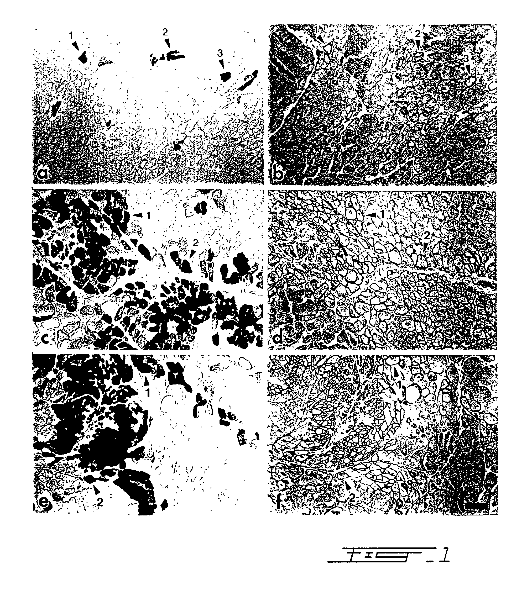
[0035] Primary myoblast cultures were established from muscle biopsies of newborn transgenic mice.sup.26. The founder mouse (TnI Lac Z1 / 29) was provided by Dr. Hasting (McGill University, Montreal, Canada) onto the CD1 background and was reproduced in our laboratory. This transgenic mouse expresses the .beta.-galactosidase gene under the control of the promoter of the quail fast skeletal muscle troponin I gene.sup.16. Blue muscle fibers are revealed in these transgenic mice following incubation with a substrate, 5-brom-4-chlor-3-indolyl-.beta.-D-galactopyronoside (X-gal) (Boehringer Mannheim Canada, Laval, Canada). Before starting myoblast cultures, it was necessary to identify transgenic newborns by X-gal staining of a small muscle biopsy because heterozygote transgenic mice were used as parents. Myogenic cells were released from skeletal muscle fragments of the transgenic newborns by serial enzyme treatments. First, a one hour di...

example 2

[0052] The above results can be extrapolated to an in vivo utility and verified in patients suffering of muscular dystrophy. The healthy donors and DMD recipients should be matched, if possible, upon their compatibility for the MHC (HLA)-class I (A, B, C) and-class II (Dr) antigens. The dystrophic patients should undertake an immunosuppressive treatment by being administered, for example, FK 506, cyclosporin, RS61443 or rapamycin. Donors' biopsy would then be treated substantially in accordance with the procedures given in Example 1 with regard to mice myoblasts. The success of the transplantation might be monitored by measuring the incidence of dystrophin-positive fibers from a biopsy obtained from the site of transplantation and by evaluating the resulting increase of muscular strength.sup.39.

example 3

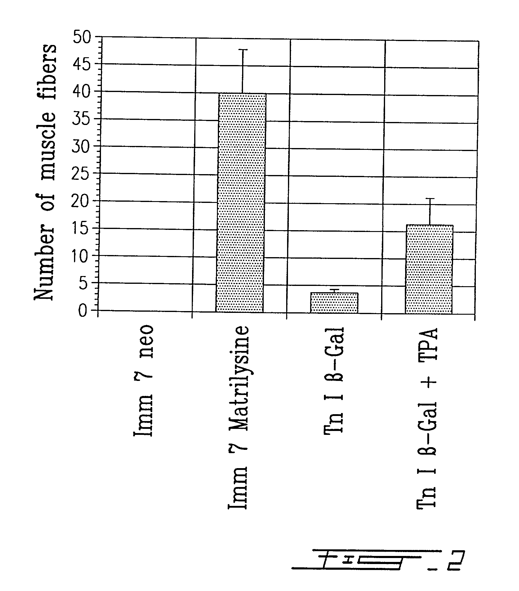
[0053] Myoblasts infected with a retrovirus expressing a beta-galactosidase gene have been cultured for four days in the presence or absence of 20.mu.g / ml concanavalin A, a lectine which stimulates the expression of metalloproteases. These myoblasts were then injected in one single site of the anterior tibialis muscle of eight mice, in order to verify the degree with which the transplanted cells are capable of migrating through the recipient tissue. After thirty days, mice were sacrificed and the muscle tissue was harvested, frozen and 10 .mu.m thick slices were mounted on slides. The presence of beta-galactosidase was revealed with X-Gal. Labelled cells were observed at a distance which is 3 to 4 fold greater in the mice muscle treated with concanavalin A. Concanavalin A is known to induce the secretion of metalloproteases by the fibroblasts present in the primary cultures. Therefore, the presence of metalloproteases in the preconditioning medium or during the transplantation is be...


PUM

PropertyMeasurementUnit
timeaaaaaaaaaa
thickaaaaaaaaaa
distance of migrationaaaaaaaaaa

Abstract

A method of pretreating healthy donor's myoblast cultures with growth or trophic factors like basic fibroblast growth factor (bFGF) and with concanavalin A on transplantation to subjects suffering of myopathy like muscular dystrophy is disclosed and claimed. Recipient muscles show a higher percentage of functional cells, a four-fold increase, demonstrated by the higher incidence of dystrophin-positive fibers, and does not require previous preconditioning of recipient muscles by irradiation or toxin administration. The recipient subjects were immunosuppressed with FK 506. When growing myoblasts with 20 mug / ml concanavalin A or 100 ng / ml TPA for two to four days, migration of donor cells in recipient tissue was increased by 3-4 fold. This suggests that, when using primary cultures, metalloproteases are secreted by fibroblasts, resulting in a greater degradation of the extracellular matrix. Both metalloproteases and bFGF appear beneficial for the success of the transplantation. The use of recombinant myoblast expressing metalloproteases is also contemplated.

Description

[0001] The present invention is a method for preconditioning healthy donor's myoblasts in vitro before transplantation thereof in compatible patients suffering of recessive myopathies, particularly of muscular dystrophy. This in vitro preconditioning improves the success of the transplantation while not requiring an in vivo preconditioning of the patient's muscle by irradiation or by administering muscular toxin.[0002] Duchenne muscular dystrophy (DMD) is a progressive disease characterized by the lack of dystrophin under the sarcolemmal membrane.sup.6,19,28,37. One possible way to introduce dystrophin in the muscle fibers of the patients to limit the degeneration is to transplant myoblasts obtained from normal subjects.sup.30,34,35. Several groups have tried myoblast transplantations to DMD patients but poor graft success was observed.sup.17,22,24,38. Even in experimental myoblast transplantation using mdx mice, an animal model of DMD.sup.10,25,29, large amount of dystrophin-positi...

Claims


Application Information

Patent Timeline
Patent Type & Authority Applications(United States)
IPC IPC(8): A61K35/12A61K38/00A61K48/00C12N5/077
CPCA61K35/12A61K38/00A61K48/00C12N5/0658C12N2501/01C12N2501/115C12N2501/59C12N2501/70C12N2510/00C12N2502/1323
Inventor TREMBLAY, JACQUES P.
Owner UNIV LAVAL
Why Eureka
Try Eureka