Mapping of differential display of proteins

a technology of differential display and protein, which is applied in the field of mapping of differential display of proteins, can solve the problems of difficult reproduction, difficult differential expression confirmation, and inability to explain the expression patterns of proteins produced from these genes

Inactive Publication Date: 2002-04-04
RGT UNIV OF MICHIGAN
View PDF1 Cites 24 Cited by
  • Summary
  • Abstract
  • Description
  • Claims
  • Application Information

AI Technical Summary

Benefits of technology

0026] The present invention relates to protein separation systems and methods capable of resolving large numbers of cellular proteins. The methods of the present invention provide protein profile maps for imaging and comparing protein expression patterns. The present invention provides alternatives to traditional separation methods for the screening of protein profiles. For example, in some embodiments of the present invention, non-porous reverse-phase HPLC is used to separate and analyze proteins as an alternative to 1-D gels. Such methods are described herein, demonstrating their effectiveness for comparing expression profiles between cells.

Problems solved by technology

While the availability of nucleic acid sequence information allows for the prediction and identification of genes, it does not explain the expression patterns of the proteins produced from these genes.
Even though these methods provide information on gene activation, there are inherent problems with them (Sturtevant, Clin. Micro. Rev., 13:408 ).
Also, bands that are differentially displayed in one trial are often difficult to reproduce in a second run and differential expression is often difficult to confirm by Northern blotting.
Gel electrophoresis methods (1-D and 2-D) have certain fundamental limitations for screening and identification of proteins from cells.
Gel electrophoresis separations are slow, where even a 1-D gel requires nearly eight hours to run with bands having sufficient resolution to study protein changes.
USA 84:6970 ; Waltham et al., Electrophoresis 18:391 ; and Henzel et al., Proc. Natl. Acad. Sci., USA 90:5011 ), the procedures remain rather labor intensive and laborious.
In addition, direct determination of the molecular weight of intact proteins from gels remains difficult, although there have been several new developments for molecular weight determination (Loo et al., Anal. Chem., 68:1910 ; Cohen and Chait, Anal. Biochem., 247:257 and Liang et al., Anal. Chem., 68:1012 ).
Another significant problem with gel electrophoresis is quantitation, where small changes in expression (plus or minus 10%) are often difficult to observe with Coomassie staining, and quantitation at any level is difficult with silver staining (Rodriguez et al, Electrophoresis 14:628 ).

Method used


Image

  • Mapping of differential display of proteins
  • Mapping of differential display of proteins
  • Mapping of differential display of proteins

Examples


example 1

MCF10 Cell Line

[0102] This example describes the properties, growth procedures, and lysis procedures of cell lines used in the following experiments. The MCF10 cell lines that were used in these experiments were obtained from spontaneously immortalized breast epithelial cells from a patient with fibrocystic disease (Soule et al., Cancer Research 50:6075 [1990]). The MCF10AT1 cell line produces xenograft lesions in immune deficient mice that resemble high risk proliferative breast disease in women. These lesions spontaneously progress to invasive carcinoma at about 25% incidence during the life of the host mouse (Miller et al., J. Natl. Cancer Inst., 85:1725 [1993]; Dawson et al, Am. Journal of Pathology 1996, 148, 313-319.). Progression of the MCF10AT1 lesions in mice is accelerated by estradiol (Shekhar et al., Int. J Oncology 13:907 [1998]). Because exposure to estrogen is a generally accepted risk factor for breast cancer development, MCF10AT1 serves as an important model to test...

example 2

Methods

[0108] This example illustrates some of the experimental methods utilized in the development of certain embodiments of the present invention.

[0109] A. Chemicals

[0110] The chemicals used in the following examples were used without prior purification. Acetone (HPLC grade) was obtained from Fisher (Fair Lawn, N.J.). Acetonitrile, guanidine hydrochloride (gu-HCl), .alpha.-cyano-4hydroxycinnamic acid (.alpha.-CHCA) trifluoroacetic acid (TFA), formic acid (FA), and octyl glucopyranoside (OCG) were from Aldrich (Milwaukee, Wis.). Trypsin was acquired from Promega (Madison, Wis.). Distilled and deionized water was obtained from Milli-Q reagent grade purification system from Millipore (Bedford, Mass.). The nitrocellulose (NC) used, Immobilin-NC pure was from Millipore.

[0111] B. HPLC

[0112] A Beckman (Fullerton, Calif.) System Gold HPLC was utilized. The pump (model 128) has a gradient solvent delivery module with built-in system controller. The detector was a programmable detector modu...

example 3

Mass Mapping of Proteins from Premalignant and Malignant Cell Lines

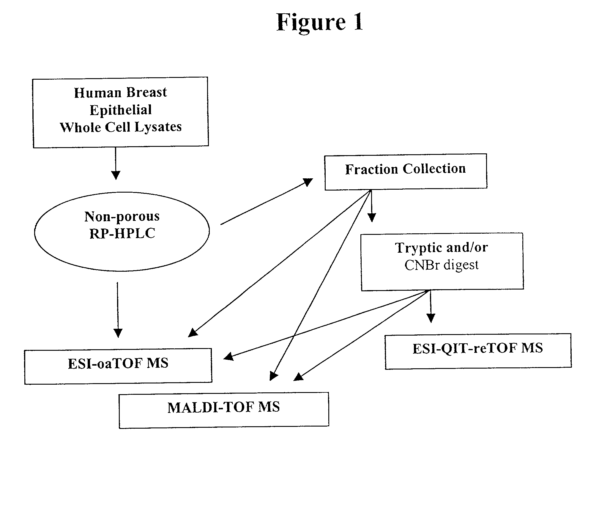
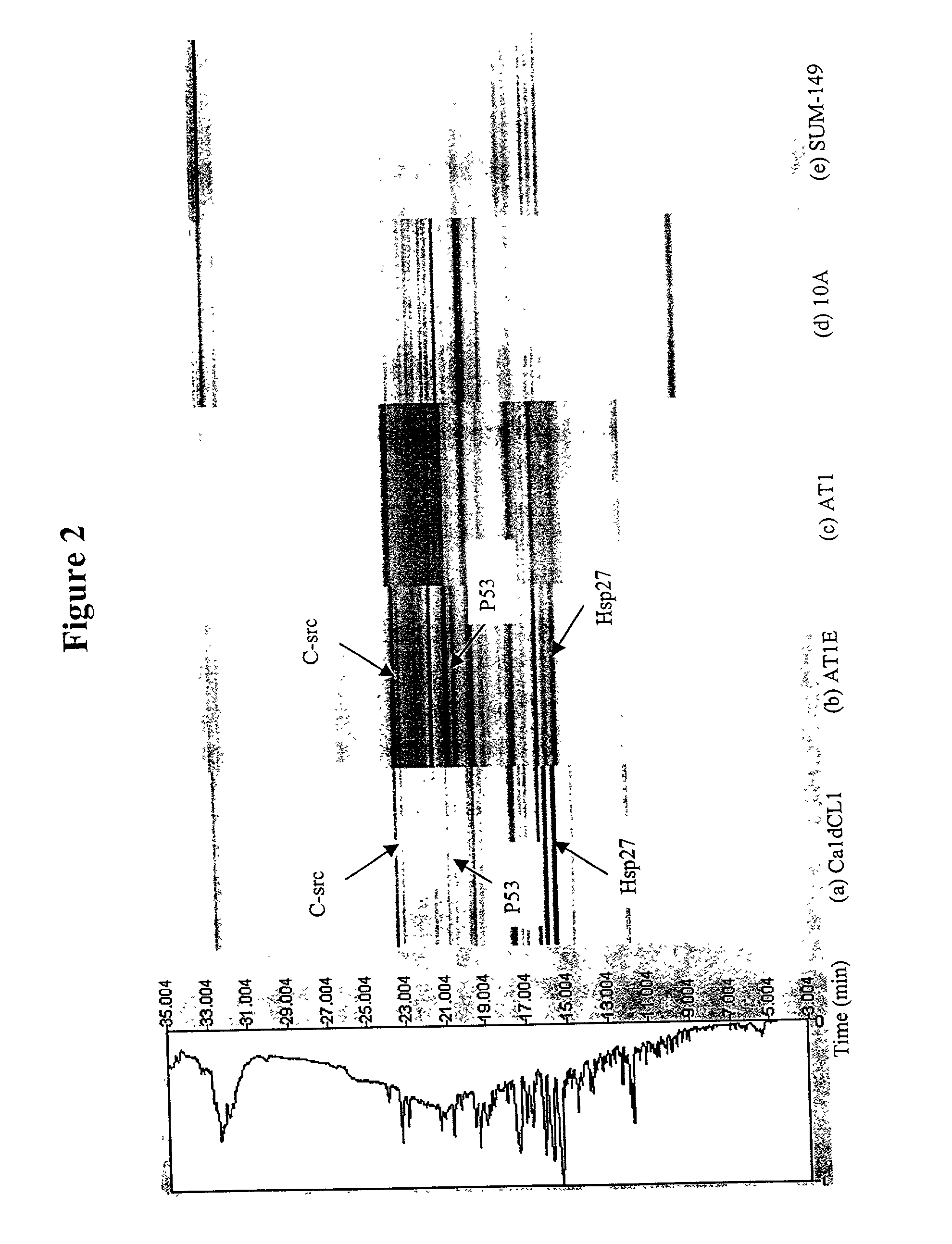
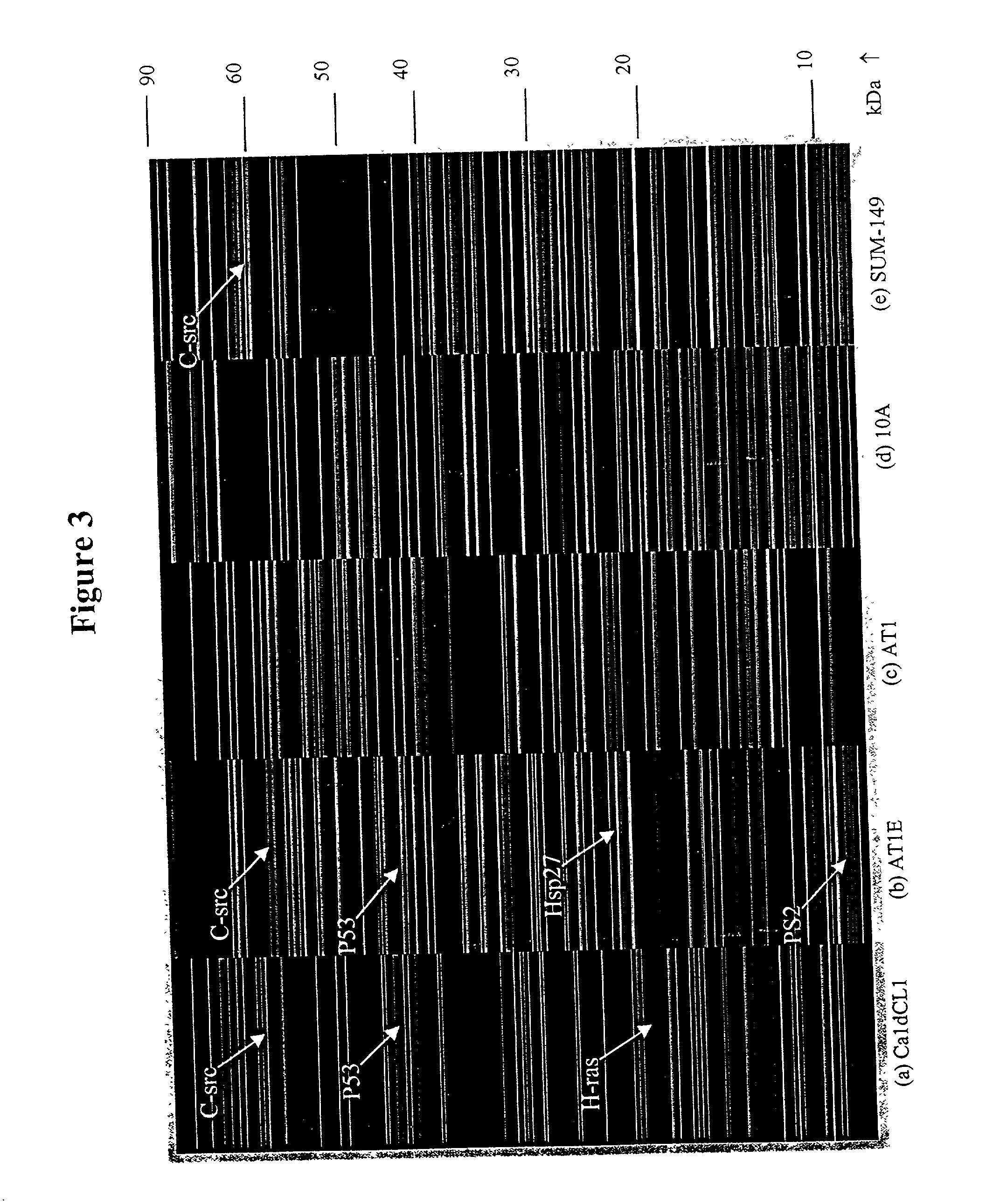
[0123] This Example describes multidimensional NP-RP-HPLC-MS analysis of human breast cell lines representing different stages of neoplastic progression. An overview of the methodology is shown in FIG. 1. The cell lines utilized included MCF10A, which is a "normal," but immortalized, cell line where the cell line keeps dividing but the phenotype is non-tumorigenic. The AT1 sample is considered a "premalignant" stage in progression. The AT1E lysate is the AT1 cell line that has been exposed to estradiol. The CaldCL1 is a highly malignant, tumorigenic cell line. These four cell lines have developed from a common precursor with essentially the same genetic background. The SUM-149 sample is a highly malignant cell line that has been developed from breast cancer tissue from a different patient and is included for comparison.

[0124] A. NP-HPLC and ESI-oa-TOF MS Analysis

[0125] An ODS2 nonporous column was used to separate th...


PUM

PropertyMeasurementUnit
mass resolutionaaaaaaaaaa

Abstract

The present invention relates to protein separation systems and methods capable of resolving and characterizing large numbers of cellular proteins. In particular, the present invention provides a novel mass mapping system and methods for the differential display of proteins. The present invention further provides novel methods for displaying differential protein expression between two samples. In particular, the present invention provides novel method of mapping differential expression of proteins in non-cancerous, pre-cancerous, and cancerous cells.

Description

[0001] This application claims priority benefit of U.S. Provisional application Ser. Nos. 60 / 180,911, filed Feb. 8, 2000, 60 / 239,325, filed Oct. 10, 2000, filed Oct. 10, 2000, 60 / 259,448, filed Jan. 3, 2001, and 60 / 259,816 filed Jan. 1, 2001, each of which is herein incorporated by reference in their entireties.[0003] The present invention relates to protein separation systems and methods capable of resolving and characterizing large numbers of cellular proteins. In particular, the present invention provides novel mass mapping systems and methods for the differential display of proteins.[0004] As the nucleic acid sequences of a number of genomes, including the human genome, become available, there is an increasing need to interpret this wealth of information. While the availability of nucleic acid sequence information allows for the prediction and identification of genes, it does not explain the expression patterns of the proteins produced from these genes. The genome does not descr...

Claims


Application Information

Patent Timeline
Patent Type & Authority Applications(United States)
IPC IPC(8): G01N27/62C07K1/36G01N30/02G01N30/46G01N30/52G01N30/72G01N30/82G01N30/86G01N30/88G01N33/68
CPCC07K1/36G01N30/02G01N30/461G01N30/7233G01N30/82G01N30/8651G01N33/6803G01N2030/8831G01N2030/027G01N2030/524G01N2030/8813B01D15/325
Inventor LUBMAN, DAVID M.CHONG, BATHSHEBA E.PARUS, STEPHEN J.
Owner RGT UNIV OF MICHIGAN
Why Eureka
Try Eureka