Directed differentiation of embryonic cells
a technology of embryonic cells and differentiation, applied in the field of directed differentiation of embryonic cells, can solve the problems of inability to determine the cell type of the embryo in vivo or in vitro, and the formation of embryoid bodies from non-human primates and humans is problemati
- Summary
- Abstract
- Description
- Claims
- Application Information
AI Technical Summary
Problems solved by technology
Method used
Image
Examples
example 1
Measuring Gene Expression Products In Differentiated Human Embryonic Stem Cells
[0058] Cell Culture:
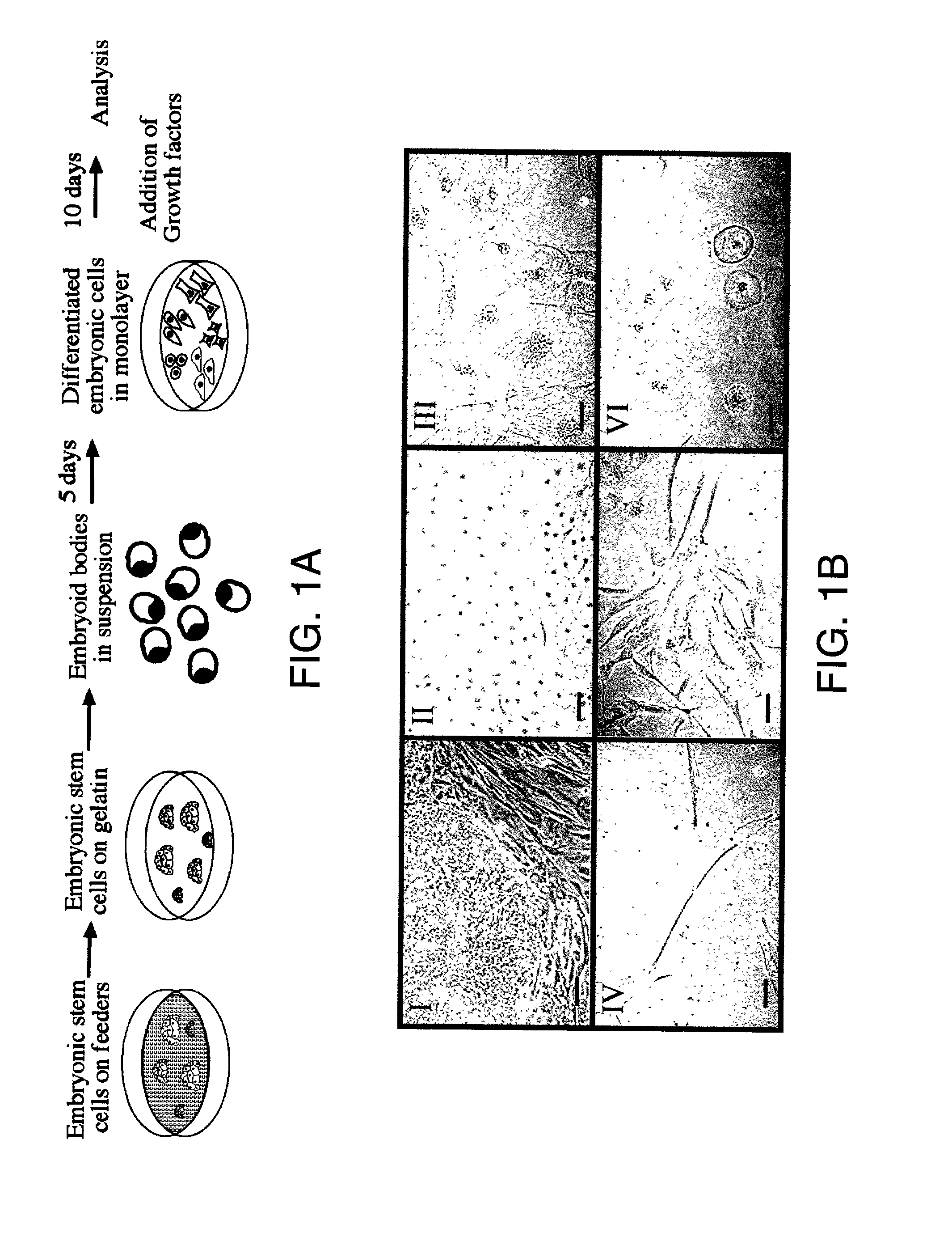
[0059] Human embryonic stem cells were grown on a feeder layer of mouse embryonic fibroblasts (MEF) and then transferred to gelatin coated plates and cultured further to reduce the number of murine cells in the culture. Differentiation into embryoid bodies (EBs) was initiated by transfer to petridishes, where the embryoid bodies remained in suspension. After 5 days, the EBs become dissociated and cells are cultured as a monolayer forming differentiated embryonic (DE) cells. This protocol allows for initial differentiation of the human embryonic stem cells as aggregates and further differentiation as a monolayer wherein cells can be exposed to exogenous growth factors. The DE cells were cultured for 10 days in the presence of 8 different growth factors: NGF, bFGF, Activin-A, TGF.beta.1, HGF, EGF, BMP4 and retinoic acid (RA) (FIG. 1-A).
[0060] Human ES cells were obtained as described in ...
example 2
Mapping Pathways Of Differentiation By Measuring The Effect Of Exogenous Growth Factors On Undifferentiated Cells
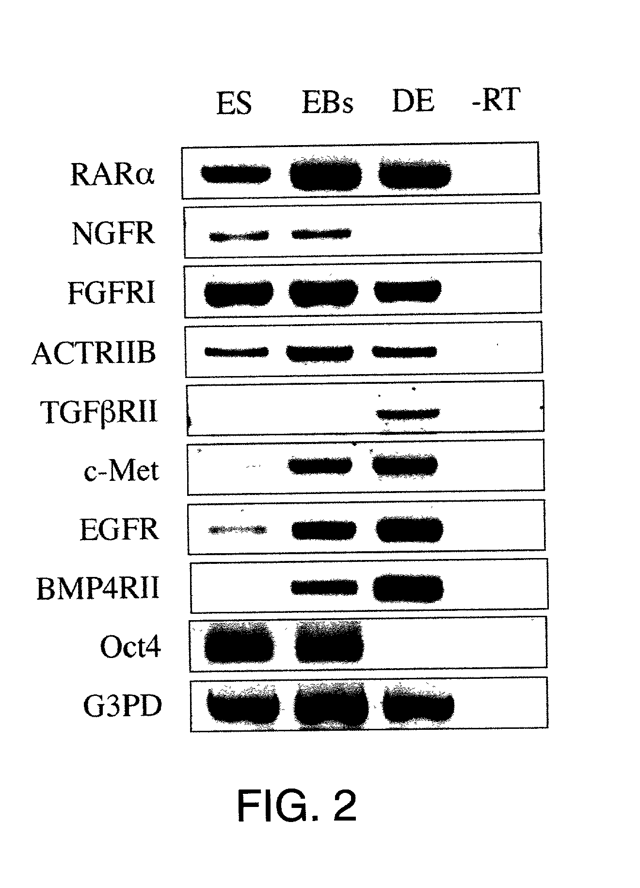
[0069] All 8 target receptors were expressed in 5 day old EBs and most were expressed in ES and DE cells (FIG. 2). Notably, four of the receptors (TGFRII, c-Met, EGFR and BMP4RII) were expressed at very low levels in human ES cells and their expression increases following culture and further differentiation. Without wishing to be limited by theory, we propose that low levels of expression may result from partial differentiation in embryonic cell cultures.
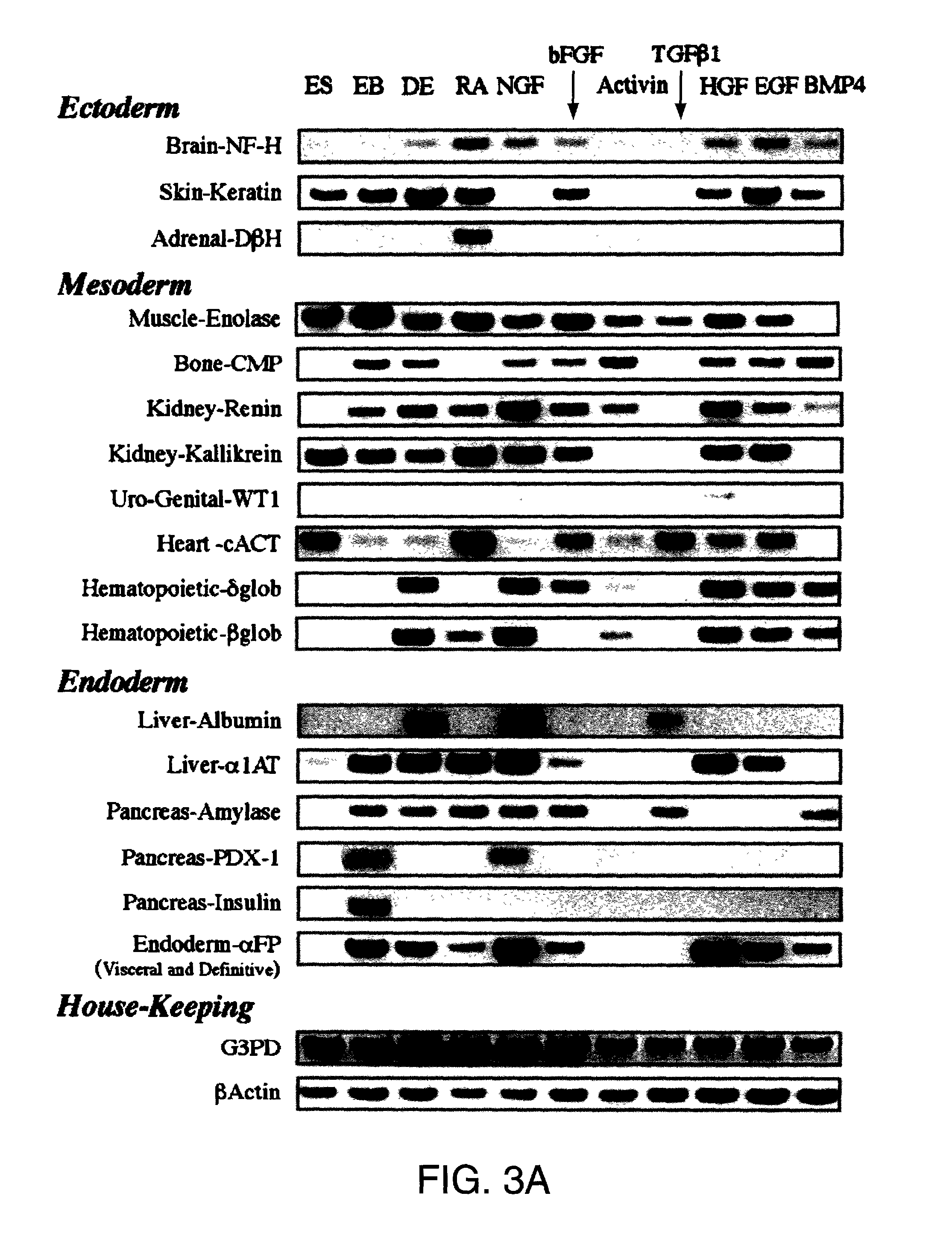
[0070] Since growth factor receptors were expressed in 5 day old EBs, we could examine the effects of the corresponding ligands on differentiation. After several days of culture on plastic and continuous exposure to growth factors, the cells acquired different morphologies (FIG. 1-B). Without growth factors, cells spontaneously differentiated into many different types of colonies, whereas the addition of growth factors pr...
example 3
Characterizing Derivative Cells From Ebs For Neuronal Differentiation
[0074] Cell Culture.
[0075] Human ES cells were grown and EBs were generated as described in Example 1. Four days following initiation of aggregation growth factors were administered: retinoic acid (Sigma): 10.sup.-7M (Bain et al (1995) or 10.sup.-6M (Bain et al., 1996); TGF.beta.1 (Sigma): 2 ng / ml (Slager et al., (1993) Dev. Genet., Vol.14, pp. 212-224.); .beta.NGF (New Biotechnology, Israel): 100 ng / ml (Wobus et al., 1988). After 21 days, EBs were plated on 5 .mu.g / cm.sup.2 collagen treated plates, either as whole EB's, or as single cells dissociated with trypsin / EDTA. The cultures were maintained for an additional week or 2 days respectively.
[0076] In-situ hybridization and immunohistochemistry was carried out according to Example 1. RT-PCR analysis. cDNA was synthesized from total RNA and PCR performed according to Example 1. Primers for GAPDH, serotonin receptors 2A and 5A (5HT2A, 5HT5A respectively) and dopami...
PUM
| Property | Measurement | Unit |
|---|---|---|
| Gene expression profile | aaaaa | aaaaa |
Abstract
Description
Claims
Application Information
 - R&D
- Intellectual Property
- Life Sciences
- Materials
- Tech Scout
- Unparalleled Data Quality
- Higher Quality Content
- 60% Fewer Hallucinations
Browse by: Latest US Patents, China's latest patents, Technical Efficacy Thesaurus, Application Domain, Technology Topic, Popular Technical Reports.
© 2025 PatSnap. All rights reserved.Legal|Privacy policy|Modern Slavery Act Transparency Statement|Sitemap|About US| Contact US: help@patsnap.com


