Helicobacter pylori antigens in blood

a technology of helicobacter pylori and antigens, which is applied in the field of methods for detecting helicobacter pylori (h . pylori) dna in blood, can solve the problems of high bacteria density, inconvenient diagnosis of h. pylori infection, and insufficient invasive tests

Inactive Publication Date: 2005-01-13
JOY BIOMEDICAL
View PDF6 Cites 5 Cited by
  • Summary
  • Abstract
  • Description
  • Claims
  • Application Information

AI Technical Summary

Benefits of technology

[0022] The target DNA sequence is further determined by running the amplified DNA in a gel electrophoresis, preferably using an agarose gel, more favorably using a 2.5% agarose gel, and staining the gel with ethidium bromide to determine the size (i.e., base pairs) of the target DNA sequence. The target DNA sequence is further determined by DNA sequencing.
[0023] The second embodiment of the present invention includes a method for detecting Helicobacter pylori (H. pylori)-specific DNA in a blood sample of a human. The method comprises the following steps: (a) obtaining a blood sample from the human; (b) extracting the DNA from the blood sample by a conventional method (such as phenol-chloroform) or a commercially available DNA extraction kit; (c) treating the extracted DNA with a denaturation agent on a solid phase support to form a denatured DNA; (d) providing an H. pylori DNA probe, which is labeled with a radioisotope or a fluorescence; and (e) detecting the presence of the H. pylori DNA by hybridizing the denatured DNA with the labeled H. pylori DNA probe by measuring the radioisotopes or fluorescence on the solid phase support. For radioisotopes, the preferred detection method is by exposing the solid phase support on an X-ray film. For fluorescence, the preferred detection method is by examining the solid phase support under a fluorescence microscope. The preferred blood sample is serum.
[0024] The H. pylori DNA probe can be prepared by any conventional genetic cloning and expression method. The preferred radioisotope for labeling the H. pylori DNA probe includes, but is not limited to, 32P, 3H, or 14C. The preferred fluorescence for labeling the H. pylori DNA probe includes, but is not limited to, a digoxigenin- and biotin-labeled DNA probe coupled with fluorescence. The denaturation agent prior to the hybridization. The preferred denaturation agent includes, but is not limited to, alkali solution (e.g., 0.1 to 1 M NaOH), elevated temperatures, organic reagents (e.g., alcohols, amides, amines, ureas, phenols and sulfoxides), or certain inorganic ions (e.g., thiocyanate and perchlorate). The preferred solid phase support is a nitrocellulose filter.

Problems solved by technology

However, H. pylori infection is not routinely diagnosed, possibly because methods of testing for H. pylori infection are not satisfactory to physicians, especially the primary care physicians (i.e. invasive biopsy test).
However, the currently available H. pylori tests, which can be categorized as invasive tests and noninvasive tests, are not completely satisfactory.
However, it requires a high density of bacteria and anything that reduces the bacterial load may produce a false-negative (Cutler, A. F., Am. J. Med.
Although the stool antigen test is reported to be satisfactory, the collection and process of the stool specimens are found to be difficult and unpleasant.
Many patients are unwilling to provide stool samples to physician due to offensive odor and lack convenient collection device.
However, these assays have not exhibited the specificity and sensitivity that are desired in serodiagnosis.
One of the problems derives from cross-reactivity.
A second problem that has been encountered in designing immunoassays for H. pylori is strain variation.
These problems preclude designing an assay around the use of a single antigen.
There are other disadvantages of using an ELISA which employs antigens to detect the presence of H. pylori antibodies in serum.
Therefore, the antigen-based ELISA does not eliminate the need for the invasive procedure.

Method used


Image

  • Helicobacter pylori antigens in blood
  • Helicobacter pylori antigens in blood

Examples


example 1

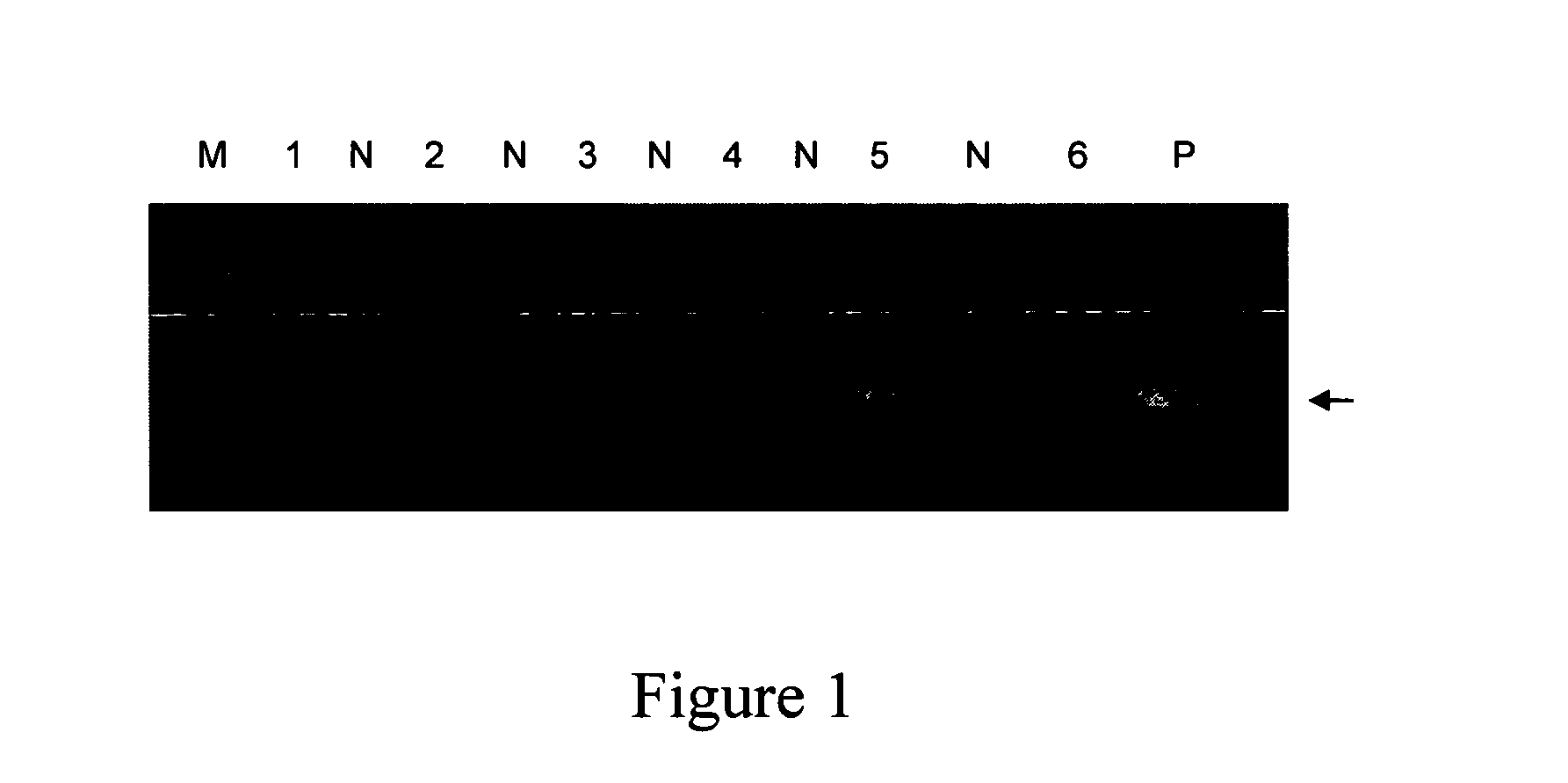
Detection of H. pylori Mr 26 Gene in Blood by PCR

[0051] Methods

[0052] Circulating DNA from 1 ml of plasma sample was extracted with a commercial kit, the QIAamp blood Mini Kit (QIAGEN). Proteinase K solution and 1 ml of Buffer AL was added to the sample. After incubation at 56° C. for about 1 hour, the sample was passed through a spin column. The majority of the high molecular weight DNA binds to the silica membrane on the spin column. The unbound impurity was washed out and DNA was eluted with 50 μl of 1 mM Tris buffer (pH 8.5). Distilled water was also subjected to DNA extraction and the elute was used as PCR negative control.

[0053]H. pylori DNA was amplified by two rounds of nested polymerase chain reaction (PCR) to detect the presence of Mr26 gene (Gene bank accession number: M55507). A pair of primers HPMr3 of SEQ ID NO:1 and HPMr4 of SEQ ID NO:2 was used in the first round of PCR and a pair of primers; and a pair of primers HPMr1 of SEQ ID NO:3 and HPMr2 of SEQ ID NO:4 was ...

example 2

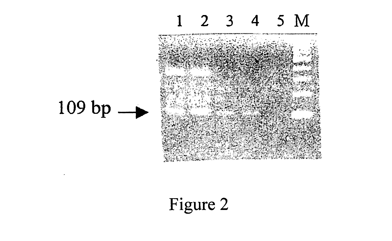
Detection of the H. pylori 16S rRNA Gene in Blood by PCR

[0058] Methods:

[0059] Circulating DNA from plasma sample of human was isolated as described in Example 1, supra. The target DNA, H. pylori 16S rRNA gene, was amplified using the same thermal cycle program as that of Example 1. A first pair of primers, U3 (SEQ ID NO:5, 5′-CAGCAGCCGCGGTAAT) and Hp1 (SEQ ID NO:6, 5′-TGGAGAGACTAAGCCTCC), was used in the first round of PCR (30 cycles); and a second pair of primers, Hp2 (SEQ ID NO:7, 5′-ATTACTGACGCTGATTGC) and Hp1 (SEQ ID NO:6, 5′-TGGAGAGACTAAGCCTCC) was used in the second round of PCR (30 cycles).

[0060] Results:

[0061] As shown in FIG. 2, the plasma sample from an H. pylori-infected human (Lane 1 and 2, duplicates) showed a DNA band after PCR which was corresponding to that of the positive control sample (Lane 3 and 4, duplicates), which used H. pylori as a template, as compared to that of the negative control (Lane 5, water), which showed no such band. The PCR product of the 16 ...

example 3

Detection of H. pylori DNA in Blood Using LCR Amplification

[0062] A ligase chain reaction (LCR) assay requires two sets of two oligonucleotides and a DNA ligase. The first set of oligonucleotides (i.e., Oligo A and Oligo B) are continuous to each other and complementary to one strand of the target DNA duplex. The second set of oligonucleotides (i.e., Oligo C and Oligo D) are complementary to the first set, and therefore occupy adjacent sites on the second strand of the target DNA. All four oligonucleotide probes can be designed according to the conserved sequence of the H. pylori strains and synthesized on an Applied Biosystems (Foster City, Calif.) oligonucleotide synthesizer and purified by PAGE. Oligo A and Oligo D can be radiolabeled at their 5′ ends by incubating for 30 minutes at 37° C. in the presence of adenosine 5′ (γ-32P) triphosphate and polynucleotide kinase in 50 mM Tris-HCl (pH 7.5), 7 mM MgCl2, and 1 mM dithiothreitol. The polynucleotide kinase can then be inactivate...


PUM

PropertyMeasurementUnit
molecular weightaaaaaaaaaa
pHaaaaaaaaaa
pHaaaaaaaaaa

Abstract

The present invention provides methods for detecting Helicobacter pylori (H. pylori) DNA and/or fragments thereof in blood. The first method involves extracting DNA from a blood sample, preferably plasma, by amplifying the DNA using a polymerase chain reaction (PCR) or a ligase chain reaction (LCR) method, and detecting a target DNA sequence in the amplified DNA. The preferred target DNA sequence comprises a Mr26 or a 16S rRNA gene or fragments thereof specific to H. pylori. The second method involves extracting DNA from a blood sample, preferably serum, by hybridizing the extracted DNA with a radioisotope or fluorescence labeled H. pylori DNA probe.

Description

RELATED APPLICATION [0001] The present application is a continuation-in-part (CIP) application which claims the priority of U.S. patent application Ser. No. 10 / 041,510, filed on Jul. 11, 2002, which is a CIP of U.S. patent application Ser. No. 09 / 572,598, filed on May 17, 2000, now abandoned, which in turn claims the priority of U.S. Provisional Patent Application Ser. No. 60 / 170,537, filed on Dec. 14, 1999. The contents of the parent applications are herein incorporated by reference.FIELD OF THE INVENTION [0002] The present invention relates to methods for detecting Helicobacter pylori (H. pylori) DNA in blood. The first method requires extracting DNA from a blood sample, preferably plasma, in humans, by amplifying the DNA using a polymerase chain reaction (PCR) or a ligase chain reaction (LCR) method, and detecting a target DNA sequence in the amplified DNA. The preferred target DNA sequence comprises a Mr26 or a 16S rRNA gene or fragments thereof specific to H. pylori. The second...

Claims


Application Information

Patent Timeline
Patent Type & Authority Applications(United States)
IPC IPC(8): G01N33/543C07K14/195C07K14/205C07K16/12C12N15/09C12Q1/68G01N21/78G01N30/88G01N33/545G01N33/553G01N33/569
CPCC07K14/205C07K16/121Y10S436/806Y10S435/975Y10S436/824G01N33/56922
Inventor YI, CHING
Owner JOY BIOMEDICAL
Why Eureka
Try Eureka