
Use of contact lens for corneal cell transplant

Inactive Publication Date: 2005-01-20
VANDERBILT UNIV
View PDF4 Cites 17 Cited by
  • Summary
  • Abstract
  • Description
  • Claims
  • Application Information

AI Technical Summary

Problems solved by technology

Current methods of corneal adult stem cell transplantation using the amniotic membrane involve a number of technically difficult steps.
First, they require the ex vivo expansion of harvested adult corneal stem cells.
The amniotic membrane is very delicate and its placement is technically complicated, requiring microsurgical instruments and specialized suture material.
If the suture fails, or the membrane is not tightly adherent, the stem cell transplantation is not successful.
Another limiting feature of this technique is that the amniotic membrane be placed with cell surface up, i.e., with the stem cells separated from the corneal surface by the amniotic membrane.
Thus, there remains a need in the art to circumvent the use of cumbersome and technically limiting protocols for the treatment of ocular conditions.

Method used


Image

  • Use of contact lens for corneal cell transplant
  • Use of contact lens for corneal cell transplant
  • Use of contact lens for corneal cell transplant

Examples


example 1

Lens Fabrication

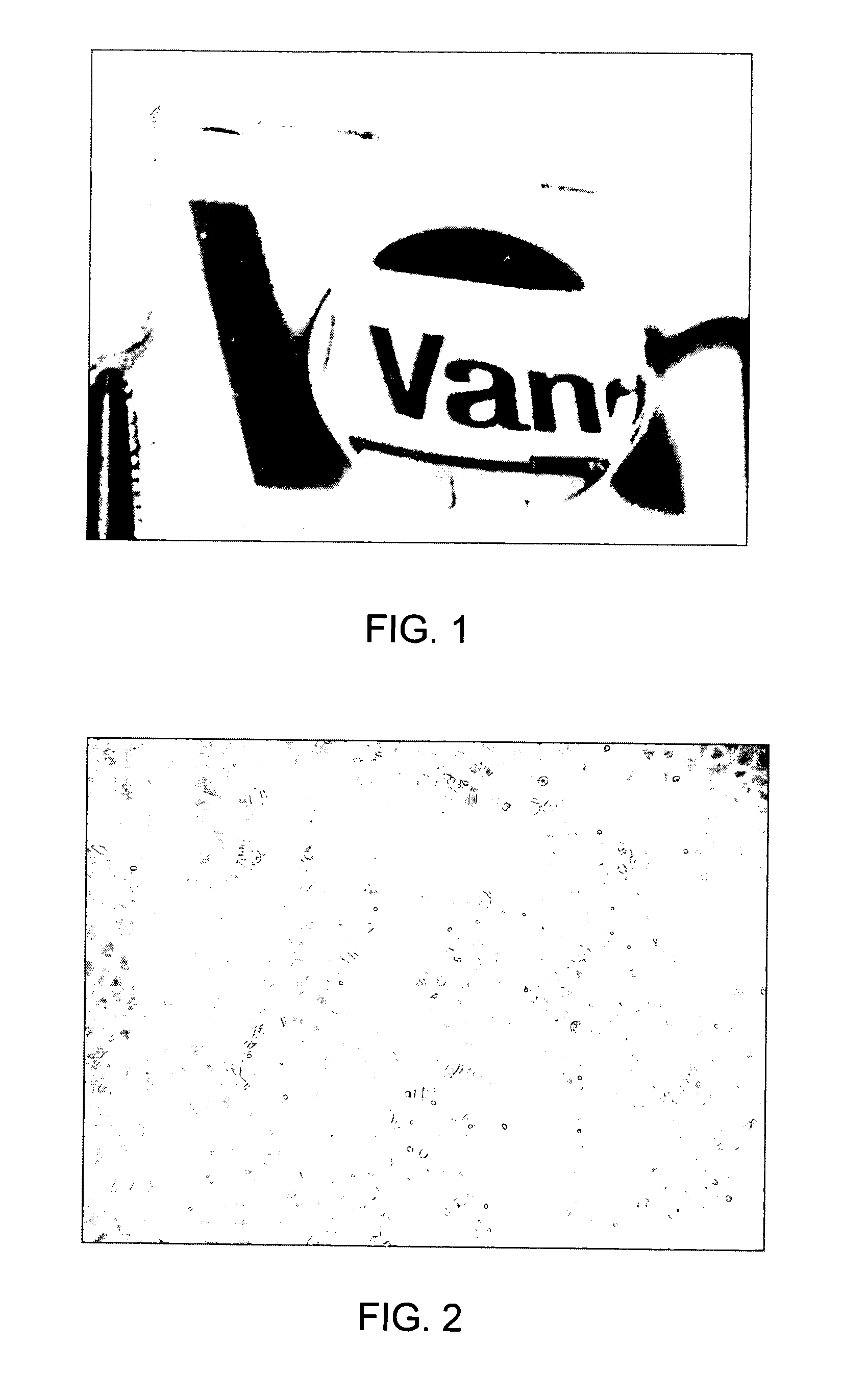
[0114]FIG. 1 shows an example of a concave molded lens fabricated from Sylgard 184 (poly (dimethyl siloxane)) (Dow Corning). The lens shown is approximately 2 cm in diameter and less than 1 mm thick and was molded using a standard drop slide following the manufacturer's instruction for the use of this material. A negative of the drop slide indentation was made and the second positive lens made by pouring Sylgard into the first negative mold and removing the material from the negative lens mold before the material had completely polymerized. The concave nature is evident from the distorted text image after the lens was placed onto newsprint.

example 2

Lens Material Supports the Growth of Epithelial Cells

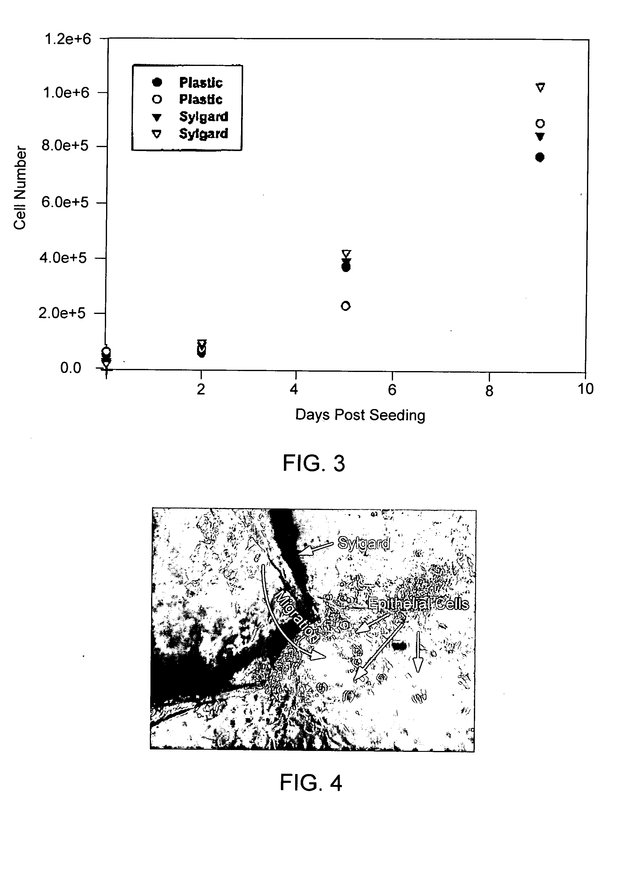
[0115] The inventors used a human corneal epithelial cell line immortalized with SV40 to test the cell growth and transfer characteristics. FIG. 2 shows an example of cultured human epithelial cells grown on the inside curvature an untreated surface of Sylgard 184. In these experiments, the molded material was autoclaved and placed into a 100 mm Petri dish with its concave side up. The mold was filled with cells plus media. The media was Gibco's defined keratinocyte media without serum supplementation. Approximately 15,000 cells (or approximately 5,000 cells per cm2) were added to the inside of the mold. Media was changed twice weekly. Cells were cultured for 7-10 days and then photographed. As shown in FIG. 2, the cells appeared confluent but patchy in some areas.

[0116] As a further test of the growth characteristics, the inventors compared the growth of Sylgard 184 with standard tissue culture plastic. In two six-well plates, ...

example 3

Cells Grown on Sylgard Migrate from Sylgard onto Tissue Culture Plastic

[0117] In these experiments, the inventors sought to demonstrate that epithelial cells will grow on Sylgard 184 but retain their ability to migrate onto another surface. This will be important in seeking to promote the transfer of cells from the extracorpeal device back onto the corneal surface. In these experiments, Sylgard 184 with epithelial cells grown on their surface were placed into a tissue culture flask cell-side down. Within 2 to 3 days, migration of the cells from the Sylgard onto the plastic was evident. FIG. 4.


PUM

PropertyMeasurementUnit
Adhesivityaaaaaaaaaa
Permeabilityaaaaaaaaaa
Gas permeabilityaaaaaaaaaa

Abstract

The present invention addresses the need for improved methods and apparatuses of transplanting cells to injured or diseased cornea. The methods disclosed provide for culture of cells on the concave surface of a contact lens, followed by application of the contact lens to the eye, thereby permitting repopulation of the corneal region by the cells. A particular use for the present invention involves the transfer of genetically altered cells.

Description

[0001] This patent application claims priority to, and incorporates by reference, U.S. provisional patent application Ser. No. 60 / 460,531 filed on Apr. 4, 2003, entitled, “Use of Contact Lens for Corneal Cell Transplant.”[0002] The government owns rights in the present invention pursuant to grant number K08 EY13592-02.BACKGROUND OF THE INVENTION [0003] 1. Field of the Invention [0004] The present invention relates generally to the fields of ocular medicine and gene therapy. More particularly, it concerns the corneal grafting using contact lens or similar biocompatible matrices. [0005] 2. Description of Related Art [0006] The cornea, along with the lens, comprise a critical focusing function for the human eye. The cornea is a thin shell only about 520 micrometers thick. Corneal epithelial cells, which cover the anterior surface of the cornea, account for about 50 micrometers of this thickness. Underlying the epithelium is a layer called Bowman's membrane which is about 20 micrometers...

Claims


Application Information

Patent Timeline
IPC IPC(8): A61F9/00A61K48/00C12N5/08
CPCA61F9/0008A61K48/0091A61K48/0075
Inventor CHANG, MIN S.HASELTON, RICK
Owner VANDERBILT UNIV
Learn More