Ultrasonic sensor garment for breast tumor

a breast cancer and ultrasonic sensor technology, applied in ultrasonic/sonic/infrasonic image/data processing, applications, tomography, etc., can solve the problems of introducing an additional degree of difficulty for a woman, abnormal tissue development has progressed past its primary stages, and the method of palpation is limited, so as to prevent the development of a fatal cancerous tumor and increase the probability of a successful treatment

Inactive Publication Date: 2005-01-27
TPG APPLIED TECH
View PDF3 Cites 35 Cited by
  • Summary
  • Abstract
  • Description
  • Claims
  • Application Information

AI Technical Summary

Benefits of technology

[0015] In one embodiment, the cancer detection system is used within the home and communicates with the doctor of the patient by way of the Internet. In another embodiment, the cancer detection system is used in a clinical setting. The cancer detection system stores all information from the periodic examinations of a particular patient and builds a chronological profile of her breast tissue development. If a localized tissue abnormality becomes apparent, proper action is taken to determine if the abnormality is the early development of a cancerous tumor. Because the cancer detection system detects abnormal tissue development in its primary stages, if a cancerous tumor is found, an immediate treatment will greatly increase the probability of a successful treatment.
[0016] The cancer detection system, in one embodiment, includes a local processing device that loads a breast examination program from a remote processing device by way of the Internet. The local processing device utilizes a sensor garment, comprised of a number of ultrasonic transceivers, to produce an ultrasonic image of the tissue ...

Problems solved by technology

This method of palpation is limited in that by the time a “lump” is large enough to be felt by the woman, abnormal tissue development has progressed past its primary stages.
Additionally, certain populations of women have naturally “lumpy” breast tissue.
This condition introduces an additional degree of difficulty for a woman attempting to detect an abnormal “lump”.
Mammograms are limited in that they are inconvenient, somewhat painful, use radiation, and produce only a “snapshot” of the organ.
Further, a shortage in radiologists has presented additional limitations to this method.
However, the most significant limitation associated with this method of breast cancer detection is the significant percentage of cancerous tumors left undetected.
However, also like mammograms, clinical examinations are inconvenient and provide only a “snapshot” of a breast.
However, ultras...

Method used


Image

  • Ultrasonic sensor garment for breast tumor
  • Ultrasonic sensor garment for breast tumor
  • Ultrasonic sensor garment for breast tumor

Examples


Embodiment Construction

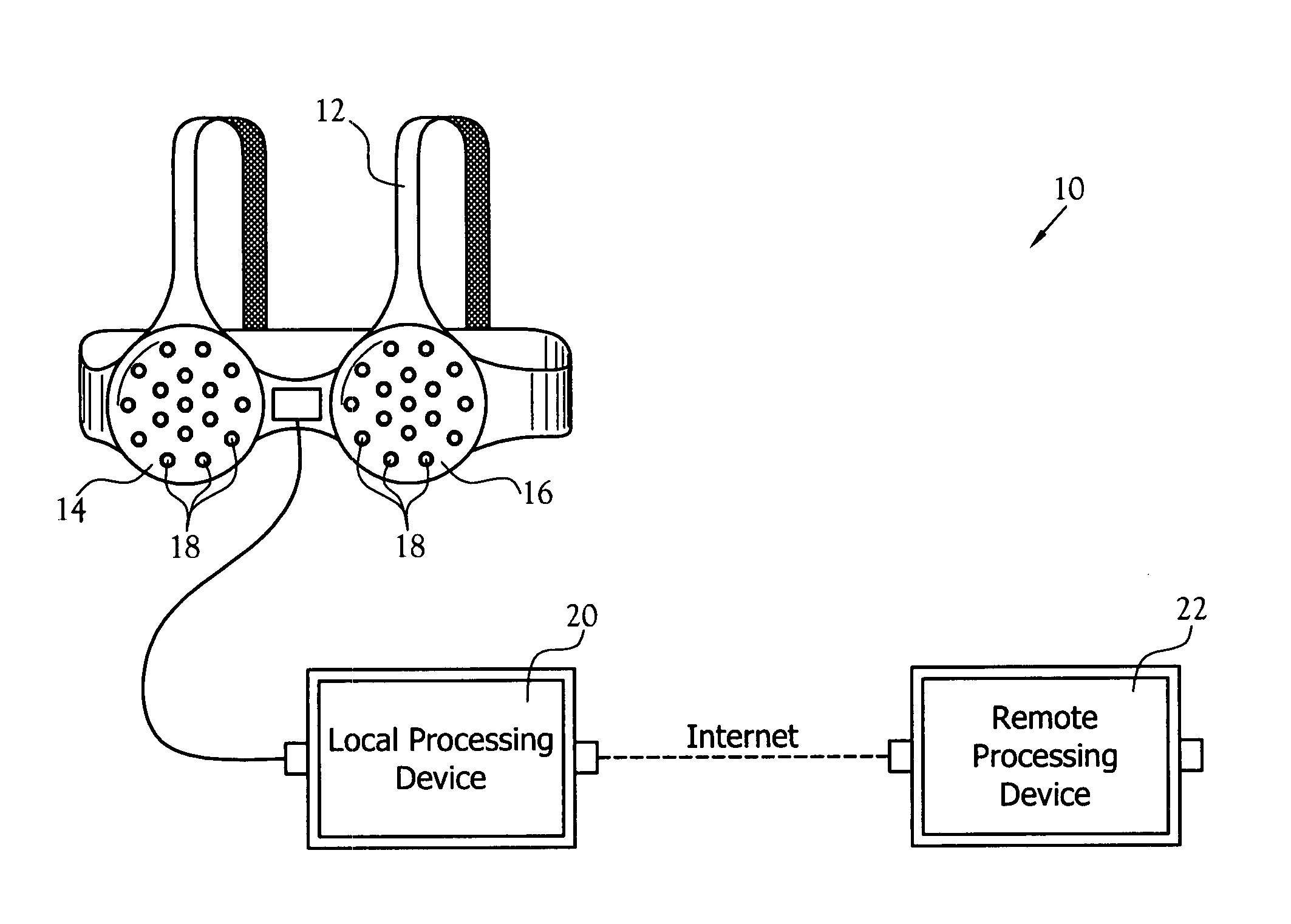
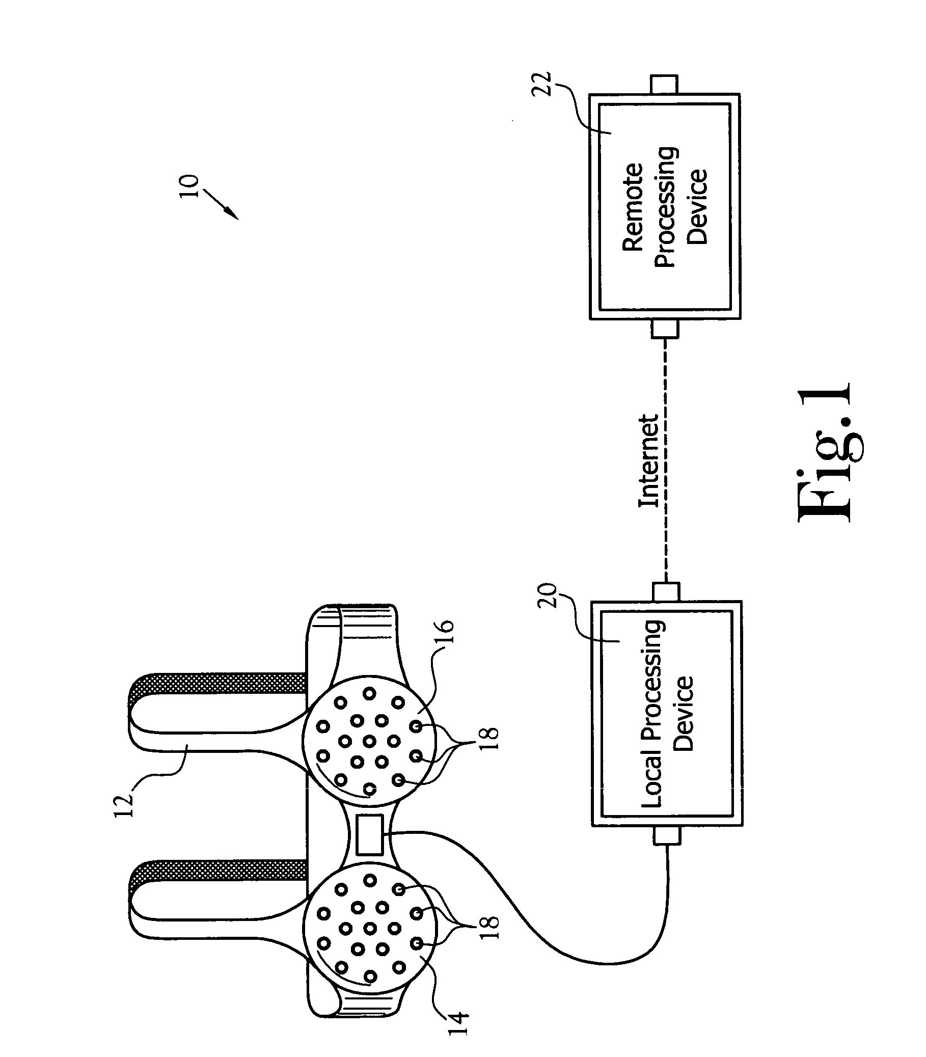
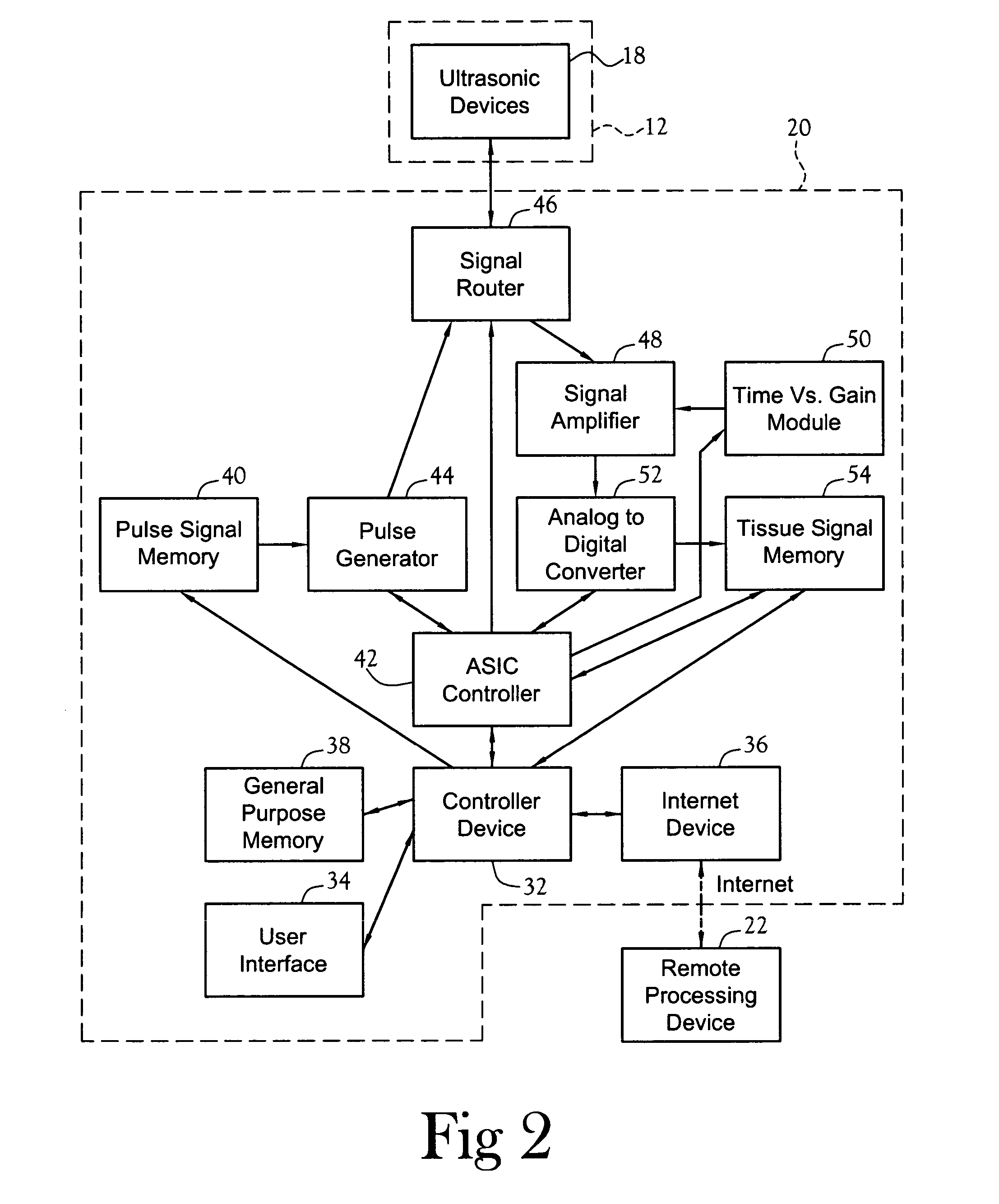
[0031] One embodiment of a cancer detection system constructed in accordance with the various features of the present invention is illustrated generally at 10 in FIG. 1. The cancer detection system 10 utilizes ultrasonic technology for gaining information regarding the tissue development of a female breast. The illustrated embodiment of the cancer detection system 10 includes a portion that is used within the home and that communicates with a remote portion by way of the Internet. The cancer detection system 10 stores all information from the periodic examinations of a particular patient and builds a chronological profile of her breast tissue development. If a localized tissue abnormality becomes apparent, proper action is taken to determine if the abnormality is the early development of a cancerous tumor. Because the cancer detection system 10 detects abnormal tissue development in its primary stages, if a cancerous tumor is found, an immediate treatment will greatly increase the p...


PUM


Abstract

A cancer detection system having a plurality of ultrasonic sensors positioned about a garment worn over at least one breast. The sensors transmit a signal that is received by the other sensors. A processor records the amplitude and time-of-flight of the received signals. The signals include both direct line-of-flight signals and reflected signals. In one embodiment, the processor performs tissue structure analysis. In another embodiment, the recorded data is sent to a remote processor for long term storage, tissue structure analysis, and/or addition to a chronological profile.

Description

CROSS-REFERENCE TO RELATED APPLICATIONS [0001] Not Applicable STATEMENT REGARDING FEDERALLY SPONSORED RESEARCH OR DEVELOPMENT [0002] Not Applicable BACKGROUND OF THE INVENTION [0003] 1. Field of Invention [0004] This invention pertains to a system for detecting cancerous tumors within a human breast. More particularly, this invention pertains to a system for ultrasonically monitoring and logging tissue development within human breasts in order to detect localized tissue abnormalities. [0005] 2. Description of the Related Art [0006] Breast cancer claims the lives of tens of thousands of women every year. Many of these victims could have survived if the cancer had been detected and treated in its primary stages. The most effective method of detecting this disease in its primary stages is regular periodic breast examinations. Currently, the three most common methods of breast examination are monthly self-examinations, annual mammograms, and clinical examinations. Monthly self-examinati...

Claims


Application Information

Patent Timeline
IPC IPC(8): A61B8/08
CPCA61B5/6804A61B8/0825A61B2562/164A61B8/4483A61B8/4227A61B8/085A61B8/4494
Inventor GLASSELL, RICHARDBURKS, BARRY L.FALTER, DIEDRE D.
Owner TPG APPLIED TECH
Why Eureka
Try Eureka