
Cell-based method and assay for measuring the infectivity and drug sensitivity of immunodeficiency virus

a cell-based method and immunodeficiency virus technology, applied in the field of genetically modified cells, can solve the problems of impeded the development of useful approaches for detecting, quantifying and analysing hiv infection of primary virus isolates, and unable to achieve cost-effectiveness

Inactive Publication Date: 2005-02-10
UAB RES FOUND
View PDF15 Cites 0 Cited by
  • Summary
  • Abstract
  • Description
  • Claims
  • Application Information

AI Technical Summary

Benefits of technology

[0017] The present invention finds utility as a method for detecting, isolating and analyzing primary HIV by infecting a cell line of the present invention with a quantity of virus and after some time measuring marker gene expression. Practicing the method of the present invention for determining immunodeficiency virus titer and conducting the presence of a drug candidate indicates the sensitivity of a given strain, type, species or genus of virus to the given candidate. The present invention affords the ability to test virus derived from blood plasma as well as cell culture.

Problems solved by technology

There is currently no cost effective, “high throughput” method for analyzing the drug resistant phenotype of primary virus isolates derived from individuals receiving antiretroviral treatment.
The sensitive detection of the virus quasispecies that comprise primary HIV isolates has proved difficult using immortalized CD4 positive cell lines.
The failure to detect infection of primary virus isolates (T-cell and macrophage tropic viruses) using immortalized cell lines has greatly impeded the development of useful approaches for detecting, quantifying and analyzing HIV infection of primary virus isolates.

Method used


Image

  • Cell-based method and assay for measuring the infectivity and drug sensitivity of immunodeficiency virus
  • Cell-based method and assay for measuring the infectivity and drug sensitivity of immunodeficiency virus
  • Cell-based method and assay for measuring the infectivity and drug sensitivity of immunodeficiency virus

Examples


example 1

Generation of Transduction Vectors for the Delivery of Marker Genes

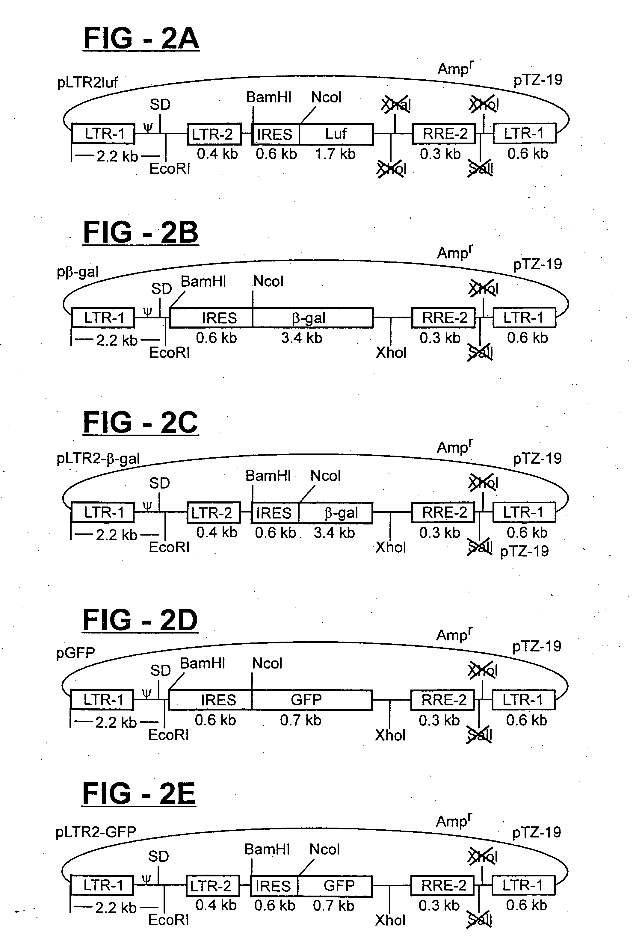
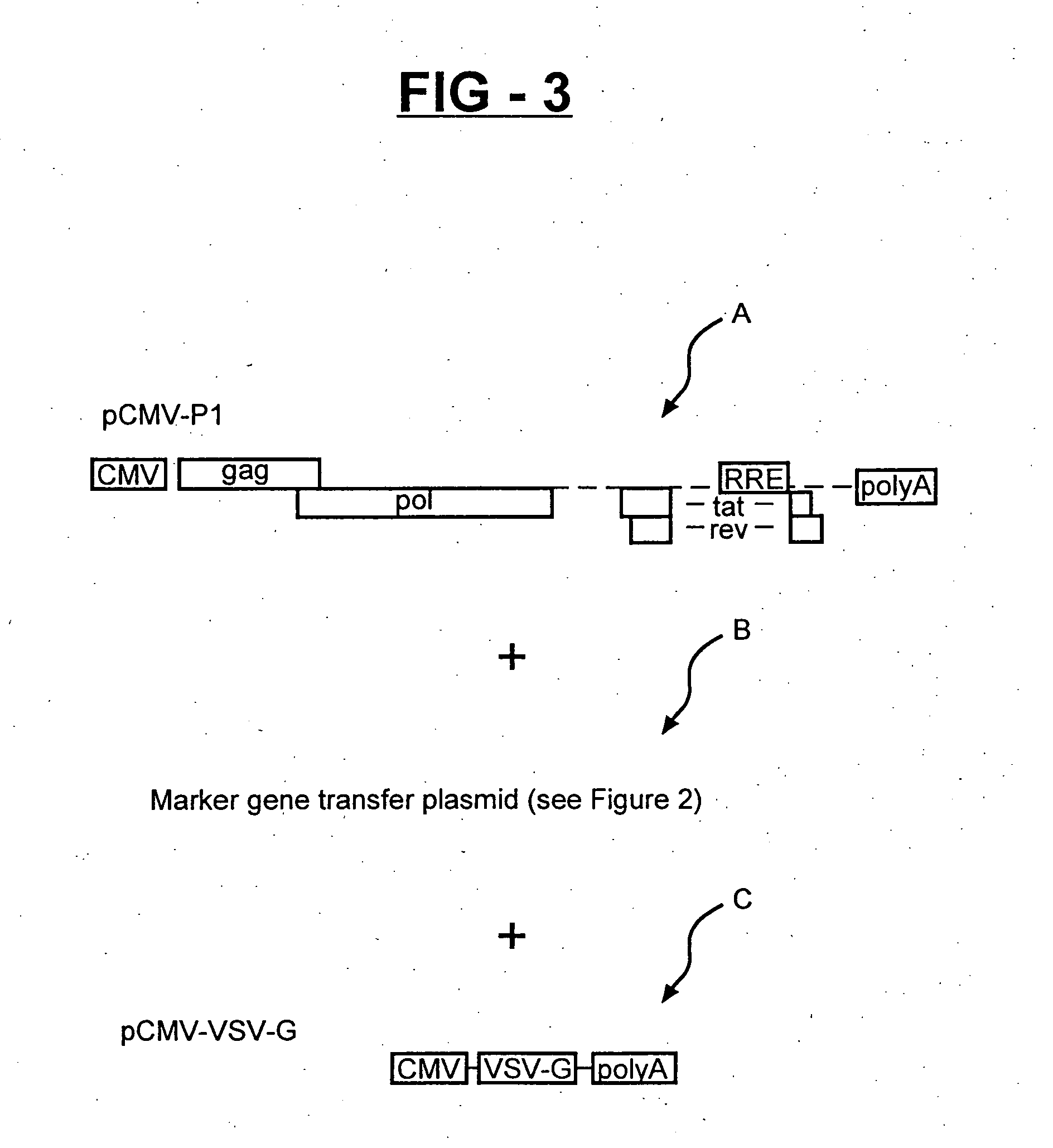
[0036] In order to generate vector stocks for transduction with the different reporter genes, the β-gal, GFP and luciferase gene transfer plasmids, including those containing, the HIV-2 LTR (shown in FIG. 2), are separately transfected into cultures of 293T cell together with a lentiviral-based packaging plasmid (pCMV-GP1), and the pCMV-VSV-G env plasmid (FIG. 3). Forty-eight hours later, the vector-containing culture supernatants are harvested, clarified by low-speed centrifugation, filtered through 0.45 micron filters, analyzed for HIV-1 p24 core antigen concentration by ELISA, aliquoted, and cryopreserved as stocks. Four serial five-fold dilutions (normalized for p24 antigen concentration) of the stocks are prepared and used to infect replica cultures of HIV-HeLa cell. The HIV-HeLa cells contained an integrated HIV-1 provirus that is defective in vpr and env, and produces the Tat and Rev protein for transactivati...

example 2

Generation of β-gal, Luciferase and GFP Indicator Cell Lines to Quantify HIV / SIV Infection

[0037] The following pairs of vector stocks (derived as described above) are used to co-transduce cultures of HeLa-CD4 cells: (a) pluf+pβ-gal, (b) pluf+pLTR2-βgal, (c) pluf+pGFP, (d) pluf+pLTR2-GFP, (e) pLTR-2-luf+pβ-gal, (f) pLTR2-luf+pLTR-2β-gal, (g) pLTR2-luf+pGFP, (h) pLTR2-luf+pLTR-2-GFP. Three days later, the cells are biologically cloned by limiting dilution in 48 well plates. Wells containing clonal cells (confirmed after initial plating by microscopy) are expanded into replica cultures. One replica culture set is infected with HIV-1 / SG3 and analyzed for marker gene expression (HFIV-1 infection provided Tat and Rev to activate marker gene expression) as described above. Expression positive cells cultures are identified, expanded and cryopreserved.

[0038] Since the expression of relatively few molecules of luciferase produces substantial luciferase activity levels, 36 non-HIV-1 infected...

example 3

Sensitive Detection of HIV-1 Primary Viruses Using β-gal and Luciferase Reporter Genes

[0039] The present invention utilizes a combination of a reporter assay system for sensitively and rapidly quantifying, infectious HIV-1 over a wide linear range with a cell line which is highly sensitive to infection with both M-tropic and T-cell tropic viruses. Transduction of the CD4-CCR5 positive J53 cell clone (Dr. David Kabat, Oregon Health Sciences University, Portland, Oreg.) with the pluf and pβ-gal expression vectors as described above. The pluf and pβ-gal transduced J53 cells (termed J53-βgal / luf) are infected with six different virus isolates (using four five-fold serial dilutions) that were unable to efficiently infect other CD4, CXCR4 expressing cell lines (P4 or Hi5) or a CD4, CXCR4 expressing cell line (MAGI) (see Table 2). Table 1 shows that all viruses, including the macrophage tropic YU2 clone, included as a control, are highly infectious in the J53β-gal / luf cell line.

[0040] To...


PUM


Abstract

Methods and reagents for the capture of primary HIV are provided. A cell line expressing CCR5, CXCR4 and CD4 receptors binds and is infected by primary HIV. The cell line contains a marker gene sequence, the marker gene sequence expressed in near linear quantities over at least two orders of magnitude in response to HIV infection. Primary HIV is amplified to create a primary virus stock through insertion of an amplicon gene into the receptor expressing cell line. HIV amplification occurs rapidly and is operative with noninfectious HIV through amplification in the presence of an infectivity complement. The present invention is useful in determining host HIV titer, drug sensitivity, HIV amplification, gene sequencing and co-receptor utilization.

Description

CROSS-REFERENCE TO RELATED APPLICATIONS [0001] This application is a continuation of U.S. application Ser. No. 09 / 719,340, filed Apr. 13, 2001; which was a national stage filing under 35 U.S.C. §371 of PCT / US99 / 14104, filed Jun. 23, 1999, which International Application published by the International Bureau in English on Dec. 29, 1999; which claims the benefit of U.S. Provisional Application No. 60 / 090,317, filed Jun. 23, 1998; all of which are herein incorporated in their entirety.FIELD OF THE INVENTION [0002] The present invention relates genetically modified cells, to an assay and methods and the usage thereof to measure the infectivity and viral resistant / sensitivity of isolate from peripheral blood mononuclear cells (PBMC) and plasma of an immunodeficiency virus. The present invention has utility in determining the HIV co-receptor usage, discovery of new drugs effective against HIV and monitoring a drug therapy protocol in order to enhance the effectiveness of drug treatment re...

Claims


Application Information

Patent Timeline
IPC IPC(8): C12N5/00C12N5/08C12N7/02C12Q1/70G01N33/50G01N33/53G01N33/569
CPCC12Q1/703G01N33/5091G01N2333/715G01N2333/70514G01N33/56988
Inventor KAPPES, JOHN C.WU, XIAOYUN
Owner UAB RES FOUND
Learn More