Multi-modality marking material and method

a multi-modality, marking technology, applied in the field of multi-modality marking materials and methods, can solve the problems of affecting the detection accuracy of markers, and affecting the safety of patients, so as to enhance the multi-modal imaging characteristics of markers

Inactive Publication Date: 2005-02-10
CARBON MEDICAL TECH
View PDF85 Cites 171 Cited by
  • Summary
  • Abstract
  • Description
  • Claims
  • Application Information

AI Technical Summary

Benefits of technology

[0032] Suitable marker materials for embodiments of the present invention include graphite, and ceramic materials such as zirconium oxide, aluminum oxide, hydroxyapatite and silicon dioxide. The marker material may also be coated with a biocompatible coating, such as carbon or a carbon resin. Carbon coated zirconium oxide may be particularly useful for embodiments of the present invention.
[0033] The marker may be sized and shaped in a variety of ways to be distinguishable from anatomical structures. In one embodiment, the marker has a major dimension between about 80 and about 10,000 microns more particularly between about 800 and

Problems solved by technology

Problems associated with the imaging techniques mentioned above include both the accurate selection and the comparison of views of identical areas in images that have been obtained at different times or by images obtained using different image modalities.
One difficulty in the use of markers for procedures utilizing multiple imaging modalities is that a marker that is detectable in and compatible with one imaging modality (e.g. X-ray) may not be detectable in or compatible with another imaging modality (e.g. MRI).
Alternatively, the marker may be detectable, but may cause substantial distortion or interference with images formed by certain imaging modalities.
Furthermore, certain markers may pose

Method used


Image

  • Multi-modality marking material and method
  • Multi-modality marking material and method
  • Multi-modality marking material and method

Examples


example

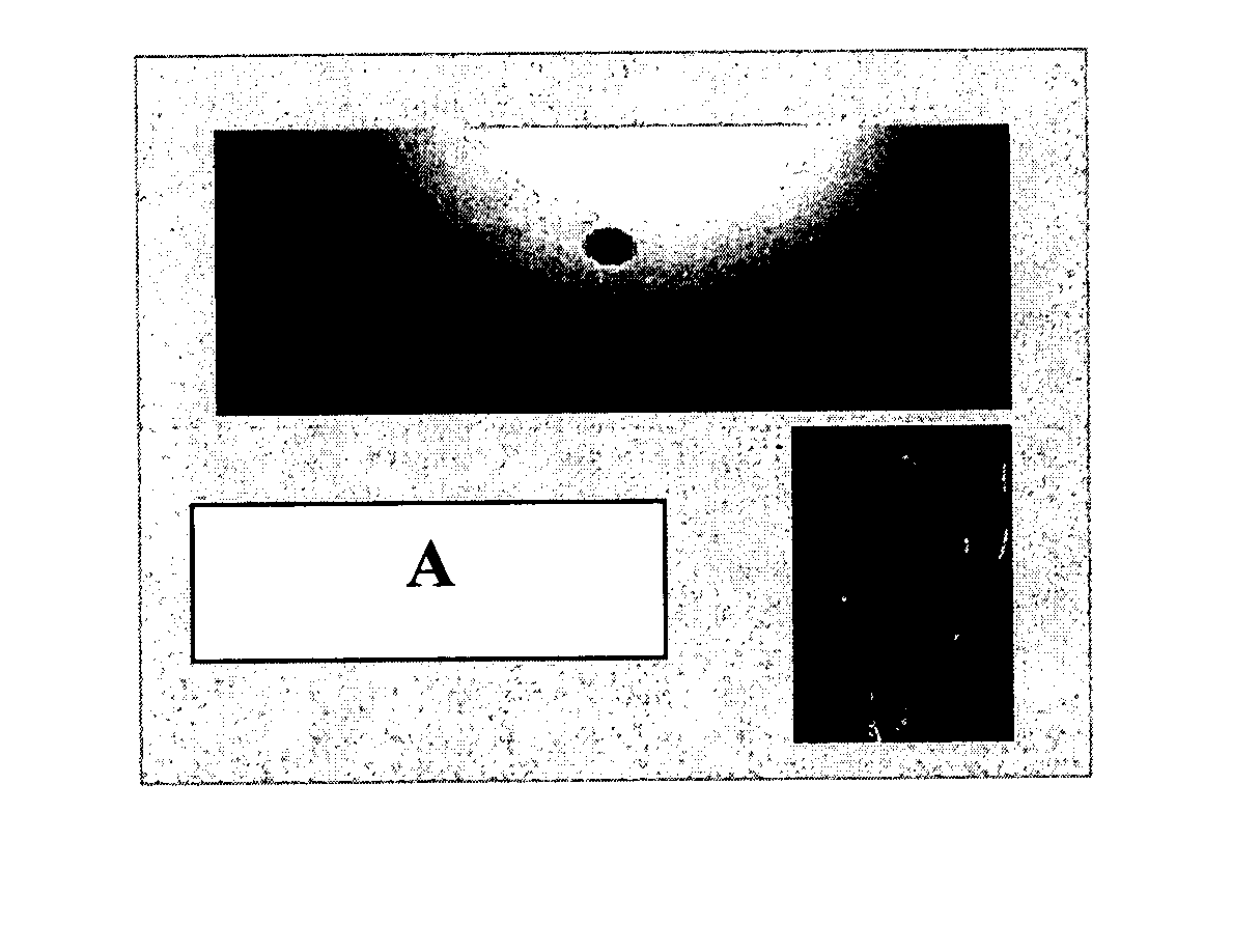
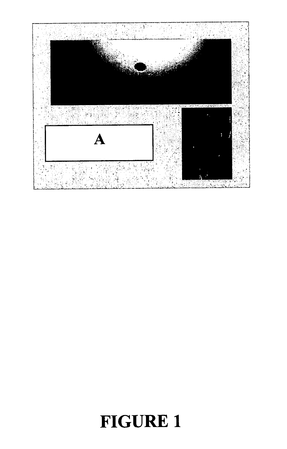
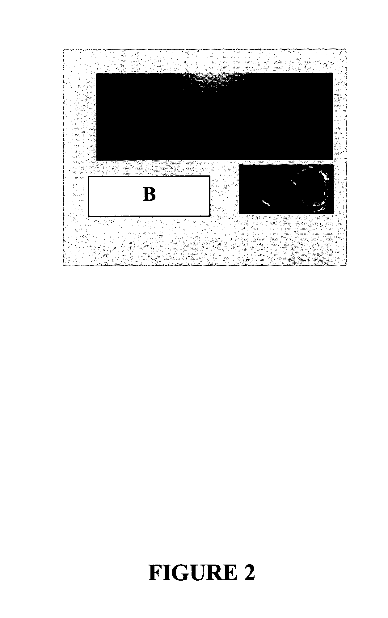
[0075] Markers A-F, each having a major dimension of 3 mm were placed 7 cm apart in a layered gelatin phantom (Knox brand flavorless gelatin, commercially available from Kraft Foods) for analysis. Markers A-C and E were composed of stainless steel alloys, marker D was composed of a titanium alloy, and Marker F was composed of a zirconium oxide substrate formed in a “dog bone” shape and coated with pyrolytic carbon.

[0076] The markers were then analyzed under ultrasound, mammography and MRI imaging modalities. The ultrasound was performed using a GE ultrasound system, mammography was performed using a Siemens system, and the MRI was performed on a Phillips 4T MRI / MRS scanner. The spatial extent of the MRI artifact was measured using a 3D FLASH image (TE / TR—6 / 17 ms, 0.4×1.7 mm resolution). Spectral distortion was measured by comparing linewidth of the water resonance from a 1 ml voxel centered on each marker, to the water linewidth measured in a control voxel containing no marker.

[00...


PUM


Abstract

The present invention provides markers and methods of using markers to identify or treat anatomical sites in a variety of medical processes, procedures and treatments. The markers of embodiments of the present invention are permanently implantable, and are detectable in and compatible with images formed by at least two imaging modalities, wherein one of the imaging modalities is a magnetic field imaging modality. Images of anatomical sites marked according to embodiments of the present invention may be formed using various imaging modalities to provide information about the anatomical sites.

Description

BACKGROUND [0001] Minimally invasive medical treatment techniques are becoming an increasingly prominent method of performing procedures for the diagnosis, treatment and / or monitoring of conditions, which were traditionally performed through an open incision. The adoption of these techniques has been made possible by the development of imaging techniques and systems that allow clinicians to obtain views or images of the anatomical features of portions of the human body. Imaging techniques and systems including computed tomographic X-ray (CT) imaging, portal film imaging devices, electronic portal imaging devices, electrical impedance tomography (EIT), nuclear medicine (NM) such as positron emission tomography (PET) and single photon emission computed tomography (SPECT), magnetic source imaging (MSI), magnetic resonance spectroscopy (MRS), laser optical imaging, magnetic resonance imaging (MRI), magnetic resonance mammography (MR mammography), electric potential tomography (EPT), bra...

Claims


Application Information

Patent Timeline
IPC IPC(8): A61B19/00
CPCA61B19/54A61B90/39
Inventor KLEIN, DEAN A.BRAZIL, JAMES D.JAEGER, THOMAS M.HALPERN, DANIEL A.GILLICK, MARK W.
Owner CARBON MEDICAL TECH
Why Eureka
Try Eureka