

Examination systems for biological samples
a biological sample and screening system technology, applied in the field of screening systems for biological samples, can solve the problems of many impediments to the proper monitoring of cultured cells, unintended changes in cell physiology, and inability to conduct around the clock monitoring
- Summary
- Abstract
- Description
- Claims
- Application Information
AI Technical Summary
Benefits of technology
Problems solved by technology
Method used
Image

Examples
example 1
[0078] Reader Module
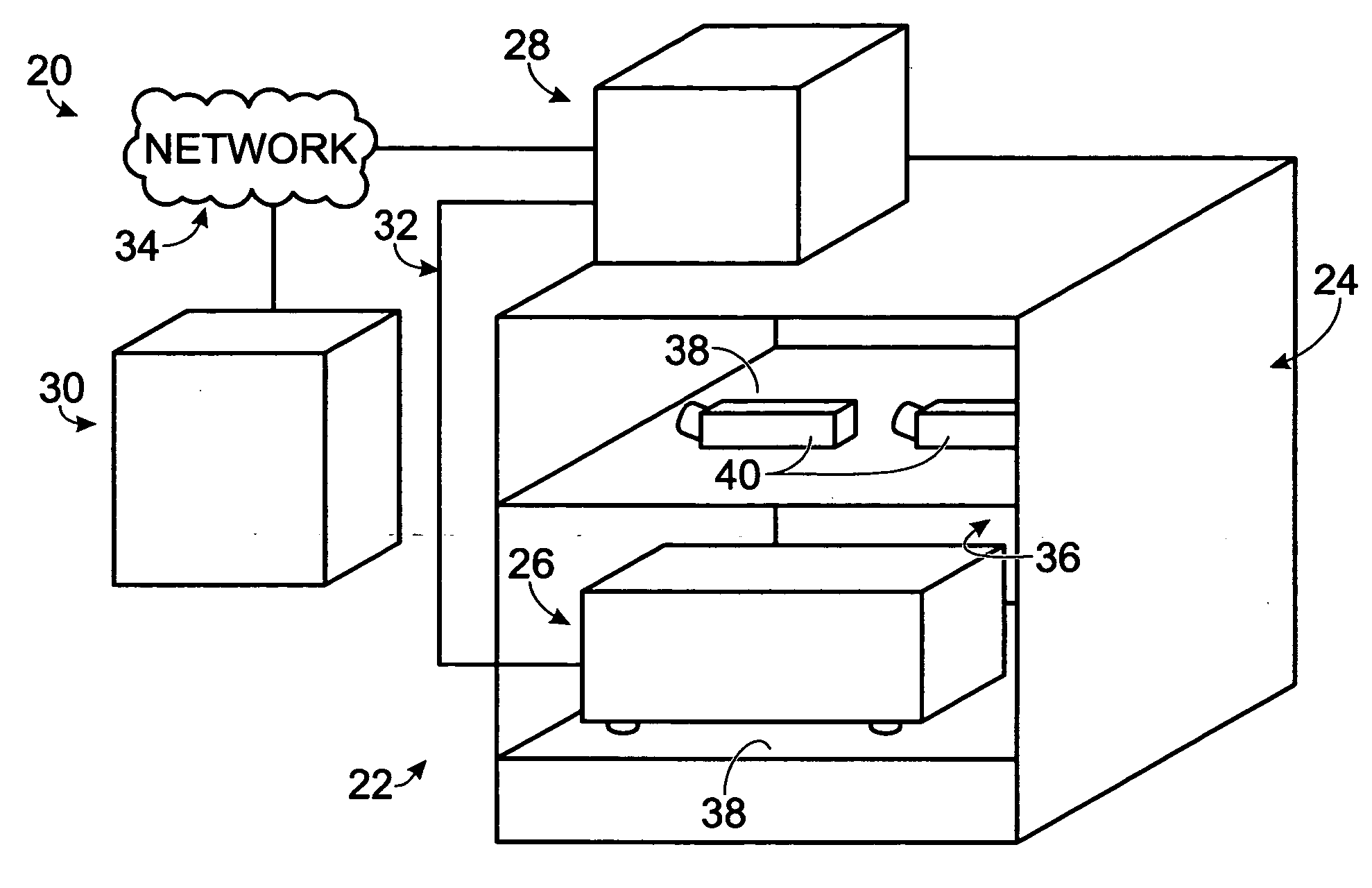
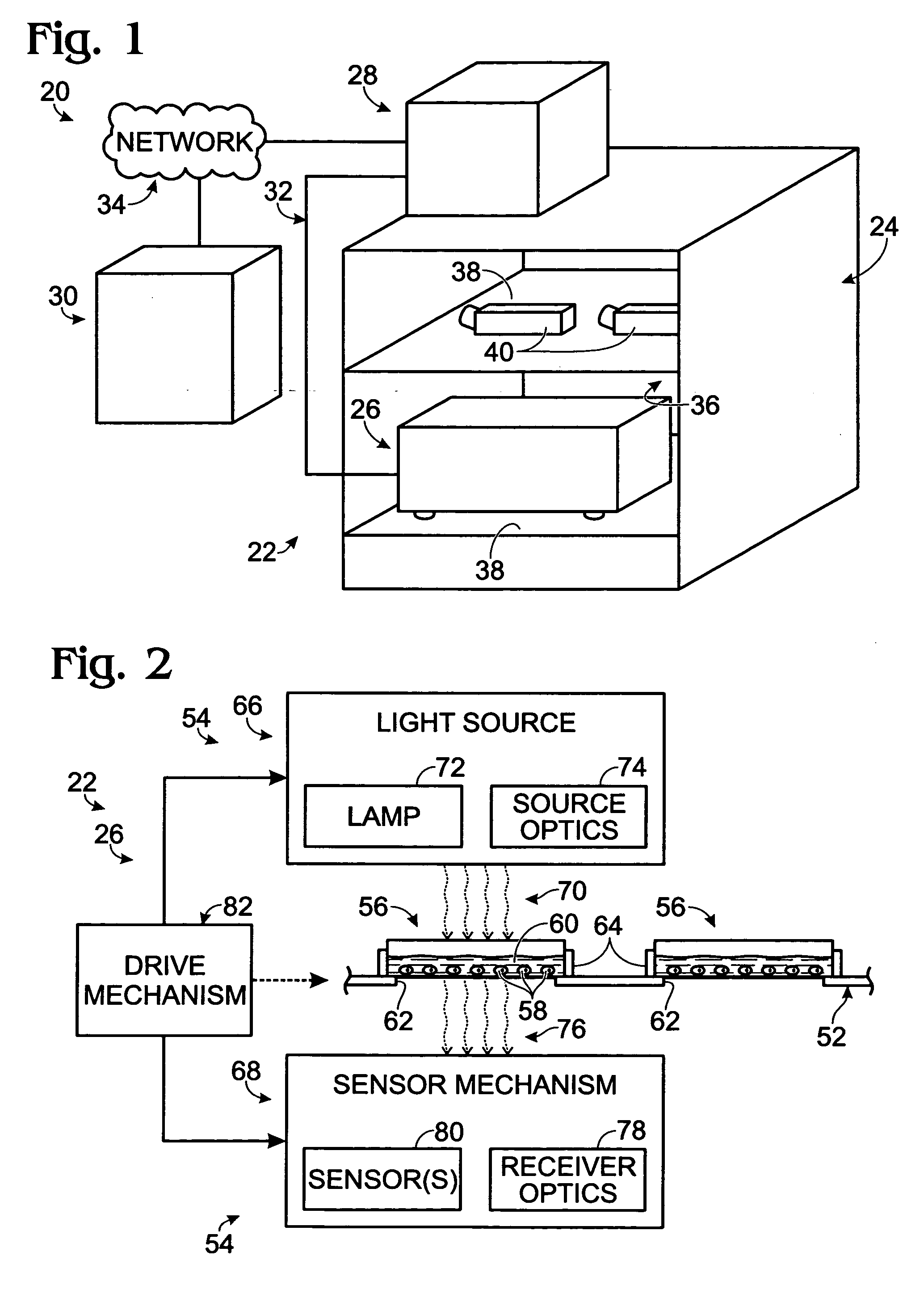
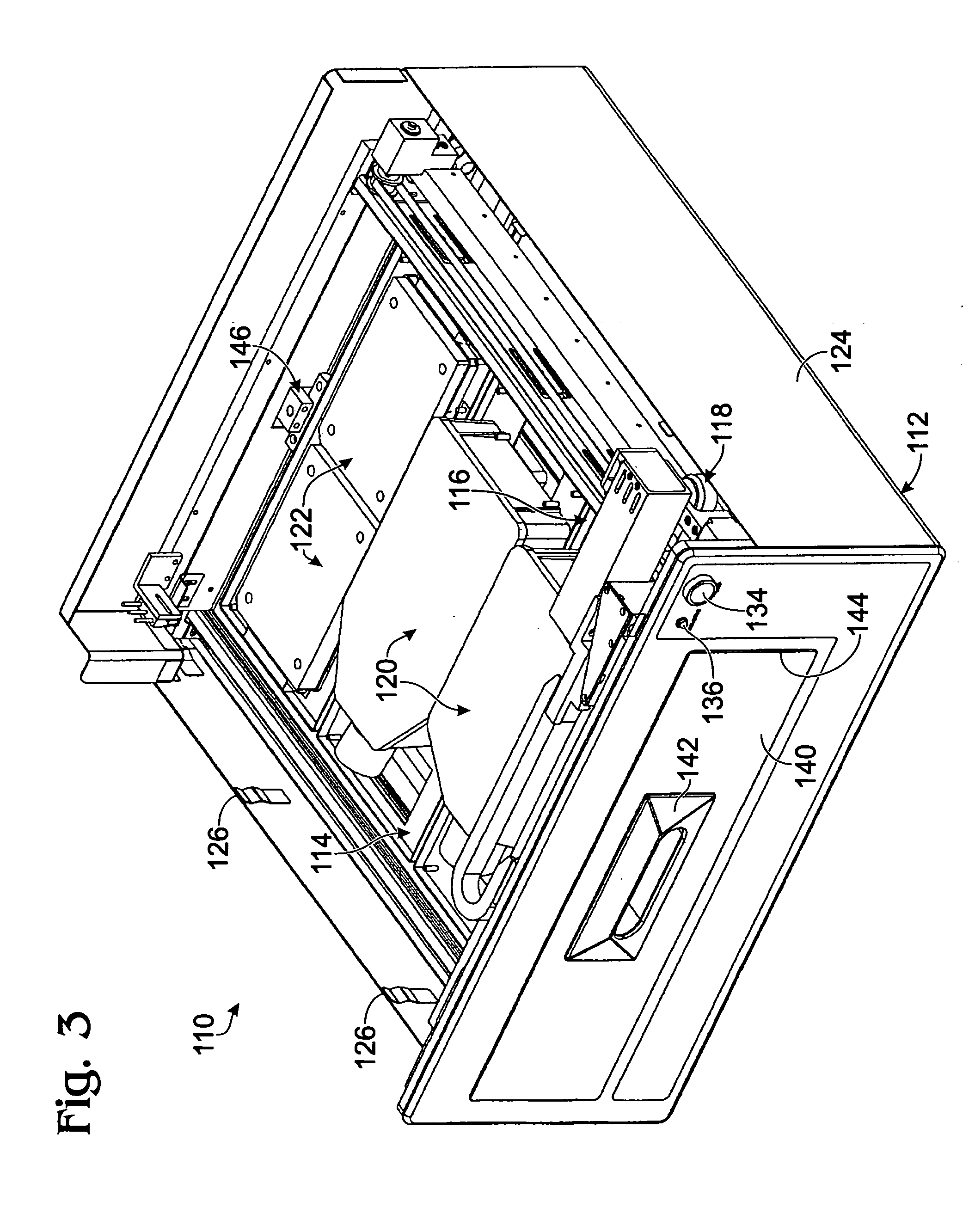
[0079] This example describes an exemplary sample reader module configured to be received in a tissue culture incubator; see FIGS. 3-9. The reader module may be included in any suitable systems for examination of biological samples.
[0080] The reader module described in this example may be used to provide microscopic examination of a plurality of plates, flasks, dishes, or microplates, among others, inside a controlled and characterized environment of a standard (commercially available) CO2 cell culture incubator. The reader module may be configured to include a microscope included in a microscope gantry that automatically scans cells in containers supported by the reader module. Because the samples remain in an incubator, kinetic images over time can be collected without the need to limit the assay time due to environmental constraints (such as unacceptable changes in temperature and / or pH of the samples). In addition, the microscope is fitted to a computer-con...
example 2
[0104] Autofocusing Features of Examination Systems
[0105] This example describes exemplary autofocusing features that may be included in examination systems, particularly for autofocusing along the Z-axis.
[0106] To keep the package size small, and the cost of the hardware at a minimum, the systems may use passive autofocusing to re-focus a microscope as it moves to different spatial locations in the sample reader. Passive auto-focusing may be image based, that is, relying on controller-based analysis of images collected by the optical system to find the optimum depth of focus for the microscope. This is in contrast to active focusing, which may rely on a special light source / detection system (e.g., a laser) to provide a means to find the optimum focus depth for the microscope. In some embodiments of the examination system, an image “sharpness metric” may be used. This sharpness metric may rely on the effective contrast achieved by the microscope in finding the bottom of a sample c...
example 3
[0108] Exemplary Interface Configurations
[0109] This example describes exemplary graphical interface configurations that may be presented to a user by a remote computing device in network communication with the local controller; see FIGS. 10-12. These graphical interface configurations may be used to perform any suitable tasks including accessing archived data and / or scheduling / configuring tasks to be performed by the local controller and / or the sample reader, among others. Exemplary graphical interface configurations may include windows termed “Tray View,”“Configuration View,” and “Flask View.”
[0110]FIG. 10 shows an exemplary screen shot of a Tray View window 310. The Tray View window may provide a pictorial snapshot of the configuration of the sample tray, as if a user were looking down on the sample tray of the sample reader. Accordingly, the Tray View window indicates the current (or a past) loaded configuration of the sample reader at a particular time, that is, the configurat...
PUM
 Abstract
Description
Claims
Application Information