Methods of cardiothoracic imaging - (MET-30)

a cardiothoracic and pulmonary vein technology, applied in the field of magnetic resonance imaging, can solve the problems of difficult imaging of thrombosis or infarct, phase errors and ghosting in the mr image, and increase the noise in the image, so as to facilitate the improvement of contrast and resolution of the stationary targ

Inactive Publication Date: 2005-03-24
KONINKLIJKE PHILIPS ELECTRONICS NV +2
View PDF66 Cites 88 Cited by
  • Summary
  • Abstract
  • Description
  • Claims
  • Application Information

AI Technical Summary

Benefits of technology

The present invention is based on the finding that stationary objects, or stationary targets as referred to herein, in an animal's body can be successfully imaged despite their location in an area subject to physiologic motion. The present inventors have found that the combination of a targeted MR contrast agent and selective timing of MR data acquisition facilitates improved contrast and resolution of the stationary target.
Accordingly, in one embodiment, the invention provides a method for determining the presence or absence of a stationary target in a bodily location of an animal. An animal can be a mammal or a bird. A mammal can be a human, dog, cat, mouse, rat, pig, or monkey. The bodily location can be the heart, lung, kidneys, great blood vessels, or the liver. A bodily location can be the myocardium, an atrium, a ventricle, a coronary artery, or a valve of the heart. The bodily location can be subject to physiologic motion. The method includes: a) administering a MRI contrast agent to said animal, with the MRI contrast agent capable of binding to the stationary target; b) allowing the MRI contrast agent to bind to the stationary target; and c) acquiring one or more MR images of the bodily location, wherein the acquisition of the one or more MR images is capable of reducing motion artifacts in the one or more MR images.
A contrast-enhancing imaging pulse sequence can include an in-flow-independent technique, which can be capable of enhancing the contrast ratio of a magnetic resonance signal of the stationary target having the MRI contrast agent bound thereto relative to a magnetic resonance signal of background blood or tissue. The background blood can be in-flowing blood. The background tissue can be fat, muscle, or tissue. An in-flow-independent technique can include an inversion-recovery prepared sequence, a saturation-recovery prepared sequence, a T2 preparation sequence, or a magnetization transfer preparation sequence.
In another embodiment, the invention provides a method for determining the presence or absence of a stationary target in a bodily location of an animal, where the bodily location is subject to physiologic motion. The method includes: a) administering a MRI contrast agent to the animal, the MRI contrast agent capable of binding to the stationary target; b) allowing the MRI contrast agent to bind to the stationary target; c) acquiring one or more MR images of the bodily location , where the acquisition of the one or more MR images is capable of reducing motion artifacts in the one or more NM images; and d) examining the one or more MR images, where the stationary target is determined to be present when a contrast-enhanced region is observed. The presence of the contrast-enhanced region or stationary target can be correlated with a pathology of the animal. The pathology can be, for example, a coronary syndrome, a coronary stent thrombosis, fibrosis of the lung, ischemic myocardial tissue, infarcted myocardial tissue, a pulmonary embolism, and a deep venous thrombosis (e.g., DVTS).

Problems solved by technology

Although MR imaging is a powerful diagnostic method for visualizing a variety of pathophysiologic and anatomic states at high resolution, a wide variety of artifacts are routinely encountered in MR images.
One type of motion artifact, view-to-view motion effects, is caused by motion that occurs between the acquisition of successive phase-encoding steps, resulting in phase errors and ghosting in the MR images.
This type of motion typically changes the amplitude and phase of the MR signal as it evolves, resulting in blurring and increased noise in the image.
Even absent such motion, imaging of a thrombus or infarct remains difficult, often due to their relative size as compared to adjacent tissue (e.g., the heart) and the lack of sufficient contrast relative to background MR signal from flowing blood and adjacent fat and tissue.

Method used


Image

  • Methods of cardiothoracic imaging - (MET-30)
  • Methods of cardiothoracic imaging - (MET-30)
  • Methods of cardiothoracic imaging - (MET-30)

Examples


example 1

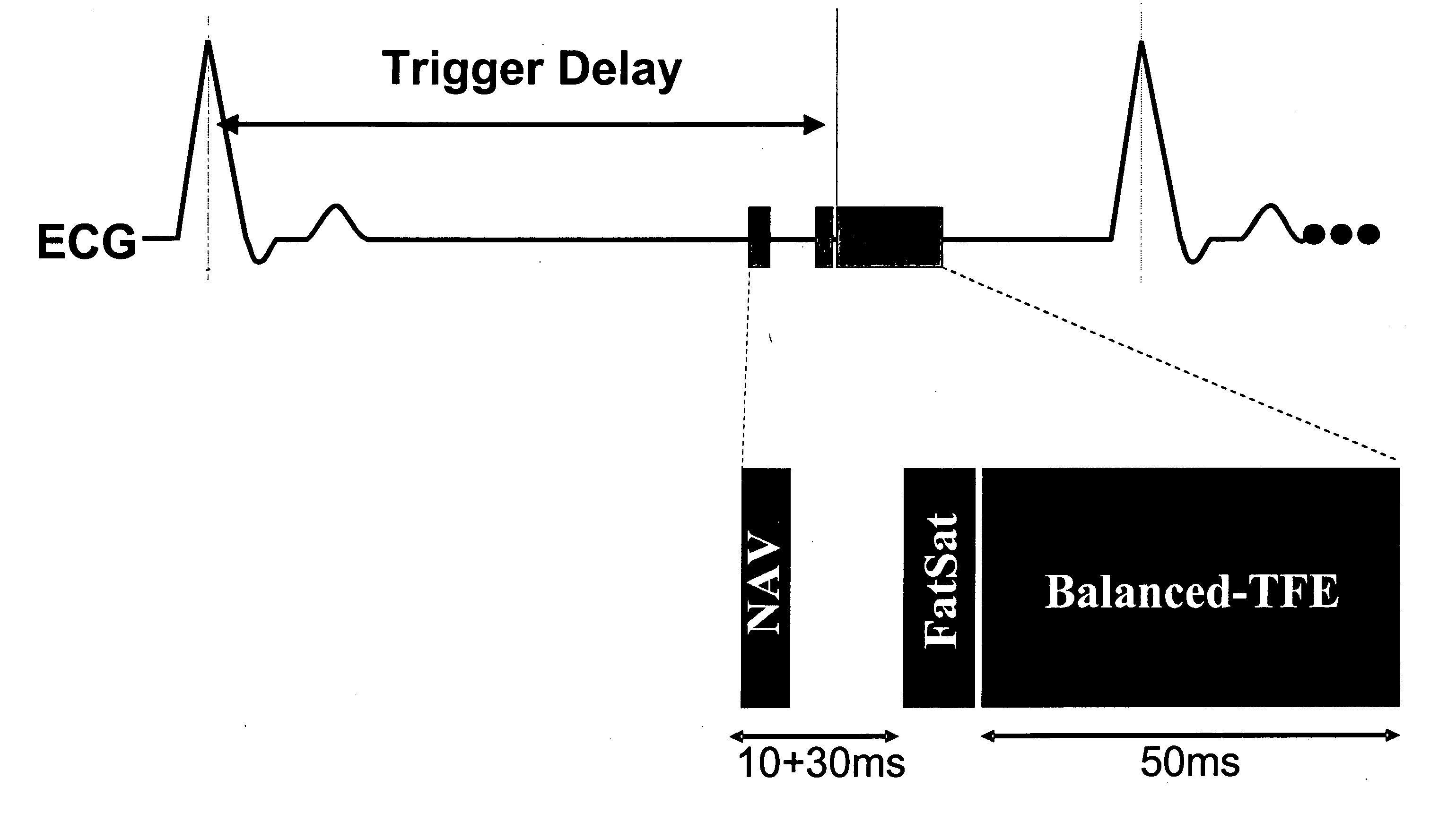
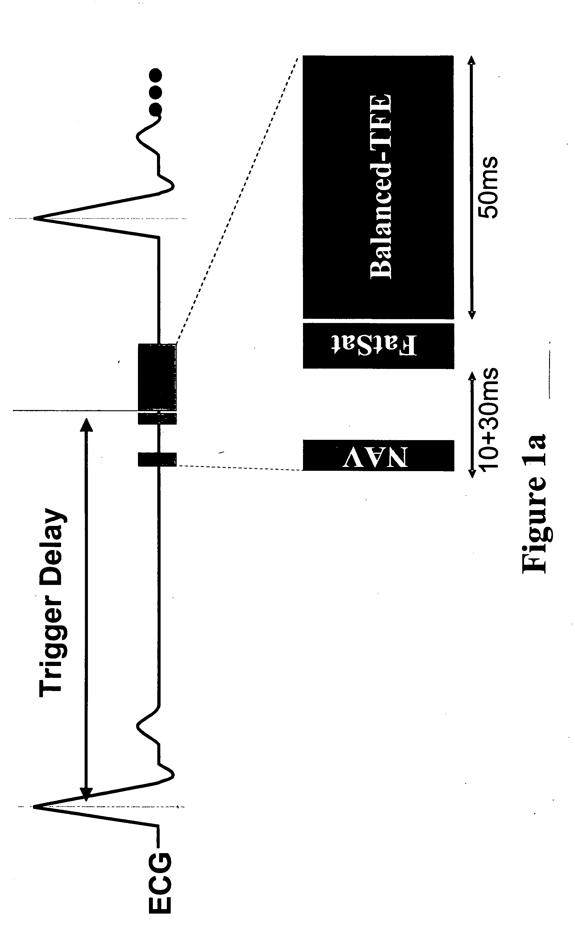
Coronary MR Angiography and Coronary Thrombus Imaging with Cardiac Triggering and Navigator Gating

Free-breathing coronary MR angiography and thrombus imaging were performed on six female domestic swine (70-80 kg) in the supine position using an interventional 1.5 T Philips Gyroscan ACS-NT short-bore MRI scanner. The MRI system was equipped with a specially shielded C-arm fluoroscopy unit (Philips Medical Systems, Best, NL), MASTER gradients (23 mT / m, 105 mT / m / ms), an advanced cardiac software patch (INCA2), and a 5-element cardiac synergy receiver coil.

Animal Protocol

After intramusculary premedication with 0.5 ml atropine and 0.2 ml azaperone / kg body weight, an aqueous solution of pentobarbital (1:3) was administered intravenously through one of the ear veins. The animals were intubated and mechanical ventilation was maintained throughout the entire experiment. A 9F sheath (Cordis, Roden, NL) was placed surgically in the right carotid artery.

MRI of Thrombi

The feasibility o...
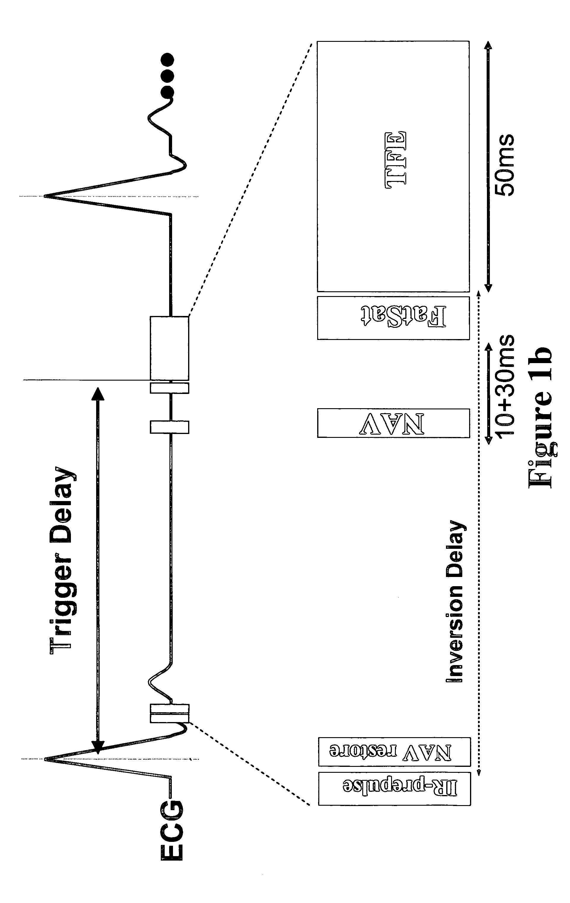
example 2

Coronary MR Angiography and Coronary Thrombus Imaging (Cardiac Triggering and Navigator Gating)—Systemic Delivery of Contrast Agent

The experiment sought to test the feasibility of direct MR imaging of acute coronary thrombosis using systemic injection of a fibrin-binding contrast agent in an in vivo swine model of coronary thrombosis. Free-breathing coronary MR angiography and thrombus imaging were performed on three female domestic swine (50 kg) in the supine position using a 1.5 T Philips Gyroscan Intera short-bore MRI scanner (Philips Medical Systems, Best, NL). The MRI system was equipped with MASTER gradients (23 mT / m, 105 mT / m / ms), an advanced cardiac software patch (R9.1.1), and a 5-element cardiac synergy receiver coil.

Animal Protocol

After intramusculary premedication with 0.5 ml atropine and 0.2 ml azaperone / kg body weight, an aqueous solution of pentobarbital (1:3) was administered intravenously through one of the ear veins. The animals were intubated and mechanical ...

example 3

Coronary MR Angiography Coronary Thrombus Imaging and Pulmonary Embolism Imaging Using Cardiac Triggering and Navigator Gating—Systemic Delivery of Contrast Agent

The differential diagnosis of acute chest pain is challenging, particularly in patients with normal ECG, and may include coronary thrombosis and / or pulmonary emboli. The aim of this study was the investigation of a fibrin-specific contrast agent (as described in Example 1) for molecular targeted imaging of coronary thrombosis and pulmonary emboli.

Animal Protocol

Coronary thrombus and pulmonary embolus MR imaging were performed on 7 healthy swine (48-52 kg BW). After premedication with 0.5 ml IM atropine, 0.2 ml IM azaperone / kg bodyweight, and 0.1 ml ketamine / kg bodyweight, an aqueous solution of pentobarbital (1:3) was administered intravenously via an ear vein as needed. The animals were intubated and mechanical ventilation was maintained throughout the entire experiment. A 9F sheath (Cordis, Roden, NL) was placed sur...


PUM

PropertyMeasurementUnit
timeaaaaaaaaaa
dissociation constantaaaaaaaaaa

Abstract

Methods for imaging stationary targets, including thrombi, are disclosed. The methods allow the imaging of stationary targets in areas of the body subject to physiologic motion.

Description

TECHNICAL FIELD This invention relates to magnetic resonance imaging, and more particularly to methods for imaging stationary targets, such as thrombi, in areas of the body subject to physiologic motion. BACKGROUND Although MR imaging is a powerful diagnostic method for visualizing a variety of pathophysiologic and anatomic states at high resolution, a wide variety of artifacts are routinely encountered in MR images. One class of artifacts, motion artifacts, is inherent in the method itself in that MR imaging equations assume stationary objects. Object motion during the acquisition of MR image data produces both blurring and ghosting in the phase-encoded direction. One type of motion artifact, view-to-view motion effects, is caused by motion that occurs between the acquisition of successive phase-encoding steps, resulting in phase errors and ghosting in the MR images. Periodic physiologic motion due to the respiratory cycle, the cardiac cycle, vascular pulsation, and CSF pulsation...

Claims


Application Information

Patent Timeline
Patent Type & Authority Applications(United States)
IPC IPC(8): A61K49/08A61K49/14
CPCA61K49/085A61K49/14A61K49/122
Inventor WIETHOFF, ANDREAPARSONS,, EDWARD C. JR.BOTNAR, RENESPUENTRUP, ELMAR
Owner KONINKLIJKE PHILIPS ELECTRONICS NV
Why Eureka
Try Eureka