Multiphoton photoacoustic spectroscopy system and method

a multi-photon and photoacoustic spectroscopy technology, applied in the field of medical imaging techniques, can solve the problems of limited diagnosis, limited application of chemical-based techniques for tumor diagnosis, and certain conditions that cannot accurately distinguish between dense healthy tissues and tumorous tissues. , to achieve the effect of non-invasive diagnosis

Inactive Publication Date: 2005-03-31
UNIV OF MARYLAND BALTIMORE COUNTY
View PDF19 Cites 111 Cited by
  • Summary
  • Abstract
  • Description
  • Claims
  • Application Information

AI Technical Summary

Benefits of technology

[0010] Another object of the present invention is to provide an improved system and method for non-inv

Problems solved by technology

Because these techniques differentiate tissues based on tissue density, they are under certain conditions unable to accurately distinguish between dense healthy tissues and tumorous tissues.
Because of this limitation in excitation wavelengths and the strong broadband absorption properties of tissues in the ultraviolet and short wavelength visible region of the electromagnetic spectrum, such diagnoses are typically lim

Method used


Image

  • Multiphoton photoacoustic spectroscopy system and method
  • Multiphoton photoacoustic spectroscopy system and method
  • Multiphoton photoacoustic spectroscopy system and method

Examples


Embodiment Construction

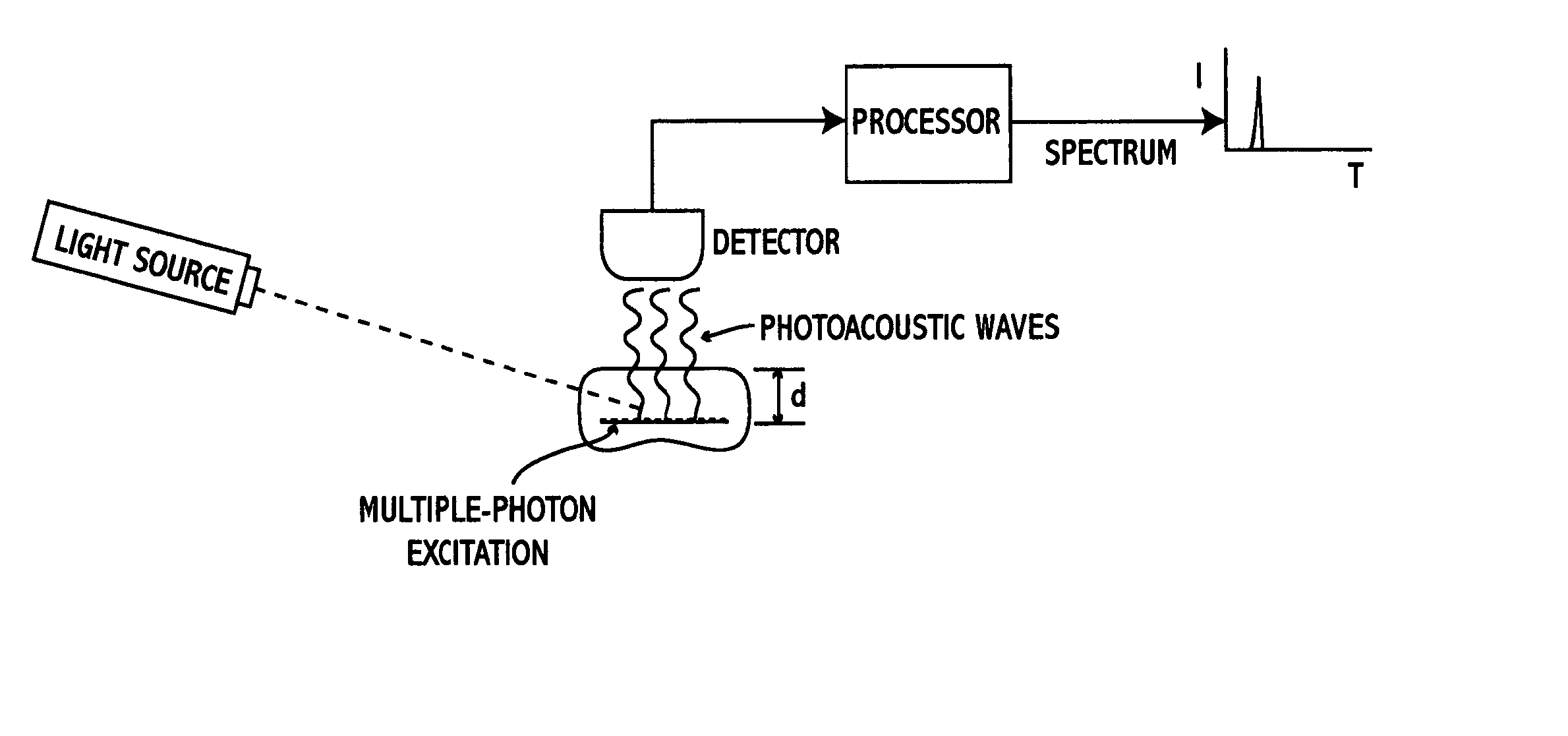
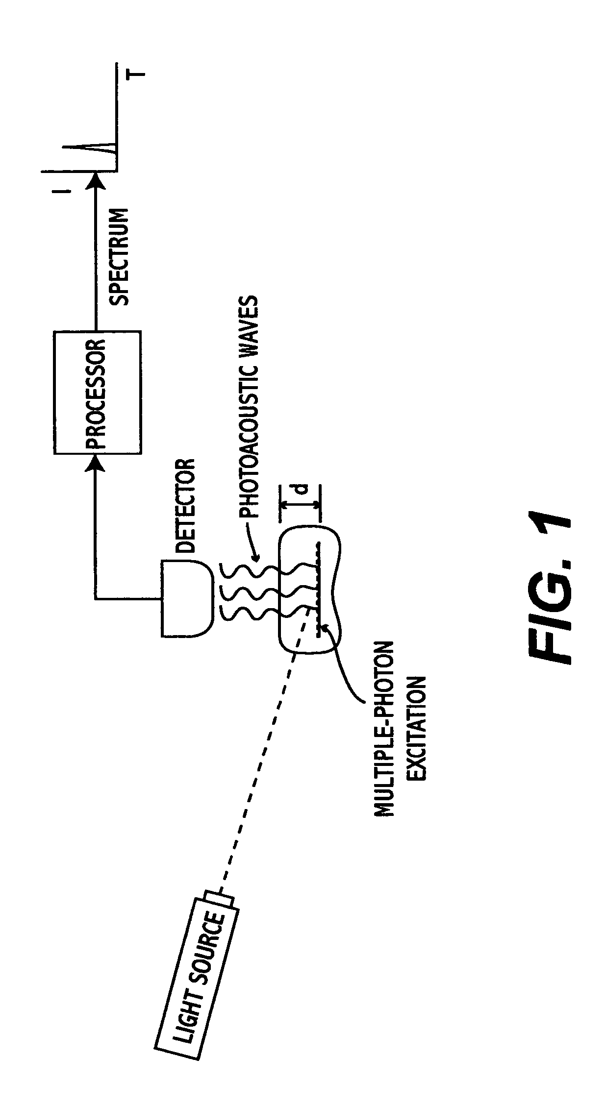
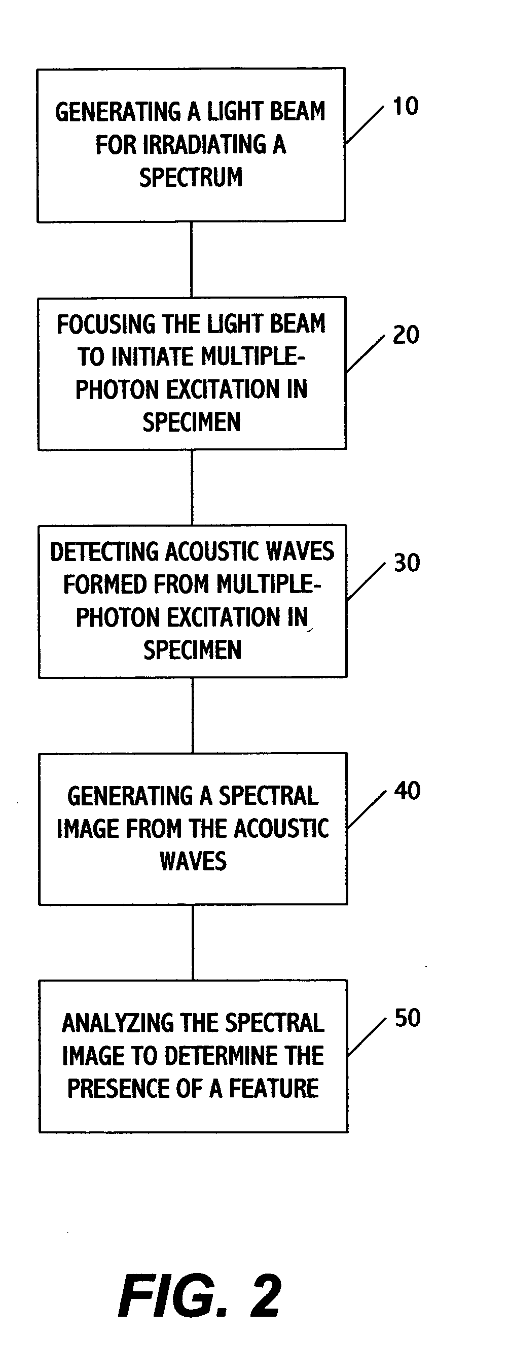
[0022]FIG. 1 shows a system for performing multiphoton photoacoustic spectroscopy (MPPAS) in accordance with one embodiment of the present invention. The system includes a light source 1, a detector 2, and a processor 3 which may be any type of general-purpose or specialized (e.g., ASIC or other chip-based) computing system. The light source is preferably a tunable high-power laser which generates a light beam within a predetermined range of wavelengths towards a specimen 5 to be analyzed. The wavelength range, laser power, or a combination of these or other parameters (e.g., objective lens power) may be selected to achieve a desired penetration depth for purposes of inducing multiple-photon excitation in the specimen accordance with the present invention, as described in greater detail below.

[0023] The detector maybe any type of transducer capable of sensing acoustic waves and converting them into electric signals. One example is a piezoelectric transducer (PZT) but those skilled ...


PUM


Abstract

A system and method for performing multispectral imaging locates features of interest in a specimen using a technique known as multiphoton photoacoustic spectroscopy. In this technique, a tunable high-power laser is used to initiate multiphoton excitation events which are then detected as an acoustic signal using a sensor such as an ultrasonic piezoelectric transducer. The transducer signal is processed to form a normalized MPPAS signal intensity which may then be used as a basis for forming a spectral image. Unlike other spectroscopies, MPPAS is able to monitor non-fluorescent species based on non-radiative relaxation of the light-absorbing species in the specimen. In addition, since the majority of energy imparted to the light-absorbing molecules is released through non-radiative pathways, sensitive measurements of even fluorescent molecules can be performed. The system and method may be applied to detect malignant cells in tissue samples although other uses are contemplated.

Description

BACKGROUND OF THE INVENTION [0001] 1. Field of the Invention [0002] This invention generally relates to medical imaging techniques, and more particularly to a system and method of performing chemical-based spectroscopy for non-invasively diagnosing anomalies or other features in specimens including but not limited to human tissue. [0003] 2. Description of the Related Art [0004] In the last couple of decades, various non-invasive diagnostic techniques such as X-ray imaging, magnetic resonance imaging (MRI), ultrasound, positron emission tomography (PET), optical coherence tomography (OCT), elastic and diffuse reflectance, photoacoustics, fluorescence, Raman scattering, etc., have been employed to diagnose malignant tumors in vivo. Depending on the method employed to differentiate between normal and tumorous tissues, these different techniques can be classified as either morphological-based or chemical-based analyses. [0005] Morphological-based methods such as X-ray, OCT, and ultrasou...

Claims


Application Information

Patent Timeline
IPC IPC(8): A61B5/00
CPCA61B5/0095A61B5/0059
Inventor CULLUM, BRIAN M.CHANDRASEKHARAN, NIRMALAMEHTA, RONAKSAHA, SOUMI
Owner UNIV OF MARYLAND BALTIMORE COUNTY
Why Eureka
Try Eureka