System and method for preparation of cells for 3D image acquisition

a technology of 3d image acquisition and system, applied in the field of specimen preparation, can solve the problems of large optical aberration, large scattering and absorption, and inability to place samples on a flat surface,

Inactive Publication Date: 2005-04-21
UNIV OF WASHINGTON +1
View PDF80 Cites 102 Cited by
  • Summary
  • Abstract
  • Description
  • Claims
  • Application Information

AI Technical Summary

Problems solved by technology

For this reason, methods such as placing the samples on a flat surface, such as a microscope slide, are not suitable, as the optical thickness of the slide and of the cover-glass (if one is used) will vary significantly as the slide is rotated by 180 degrees about one of its lateral dimensions.
Unfortunately, variations from multiple perspectives when viewing a slide can produce large optical aberrations, as well as a large degree of scattering and absorption.
Such large optical aberrations may render the projections taken unusable, especially if taken from a perspective close to the plane of the slide.
Because of the small sizes involved, it may prove difficult to insert the cells into, for example, a small capillary tube.
Glass capillaries tend to be brittle, and hence easily broken.
If the sample to be examined includes a large number of cells, strung out along a long length of glass capillary tubing, then their storage and transport can be very difficult.
The alternative method of using a large number of short tubing segments is equally unappealing.
Further, if the mechanism for insertion makes use of capillary rise, then the method may be subject to constraints imposed

Method used


Image

  • System and method for preparation of cells for 3D image acquisition
  • System and method for preparation of cells for 3D image acquisition
  • System and method for preparation of cells for 3D image acquisition

Examples


example method

for Cell Preparation for Buccal Scrapes in 3-D Visualization

General Sample Collection

[0077] An alternate embodiment of the method of the invention for buccal scrapes is described hereinbelow. Scrapings of the internal aspects of the oral cavity, that is, buccal surfaces of the cheek, are obtained as by using a plastic scraper or the like. Care should be taken to avoid abrading so vigorously as to cause bleeding. After scraping both left and right buccal surfaces, the scraper is placed into a container of isotonic solution for preservation of cytology specimens and for the liquefication of mucus. Mucoliquefying transport fluid for the collection and transport of fresh cytological specimens such as Mucolexx® available from Thermo Electric Corp., Pittsburgh, Pa., US, is used to cover the area containing the scrapings. The scraper is agitated very briskly for 20-30 seconds to dislodge any cellular material, then the scraper is removed and discarded.

[0078] The following steps are then...


PUM


Abstract

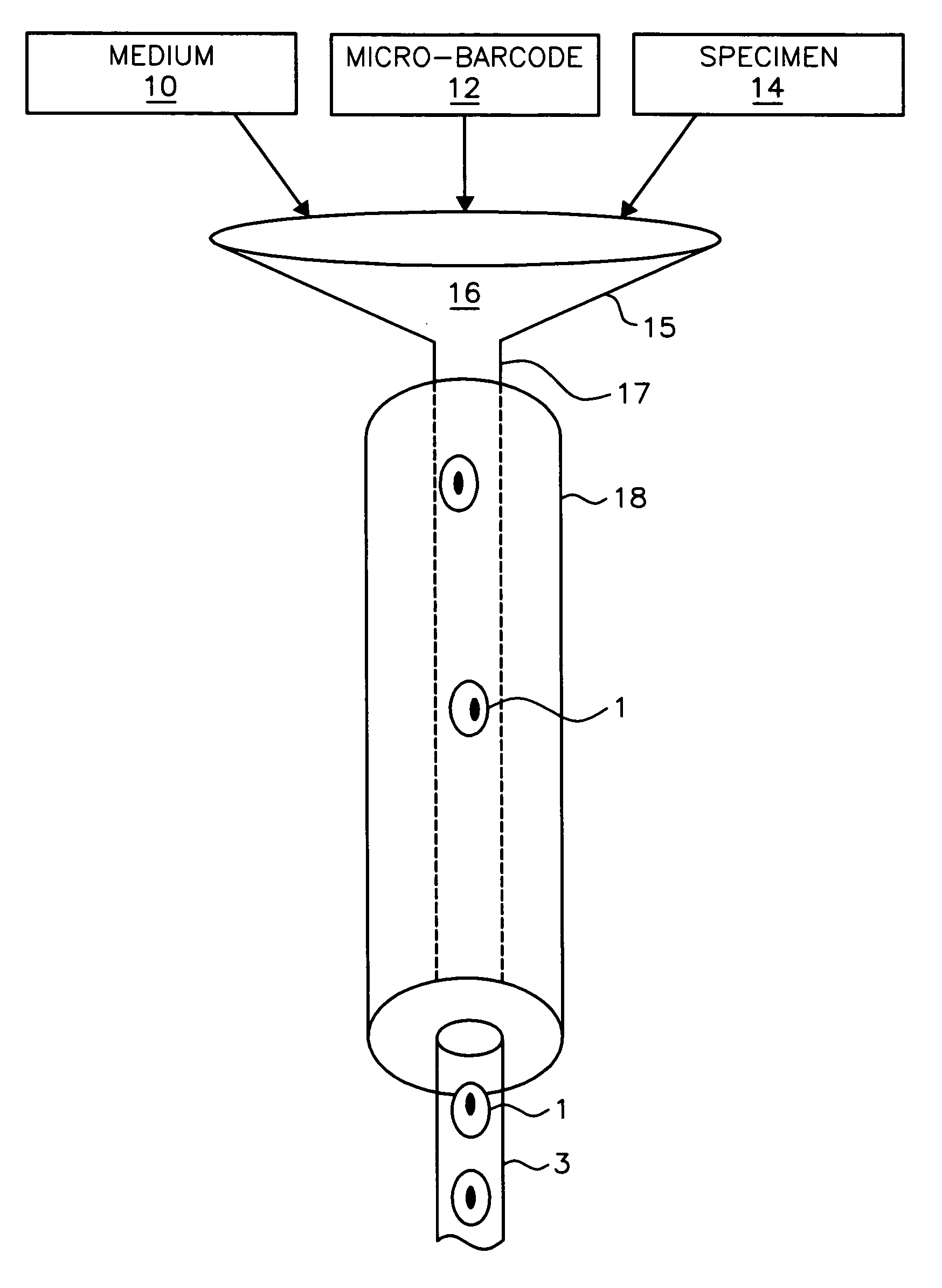
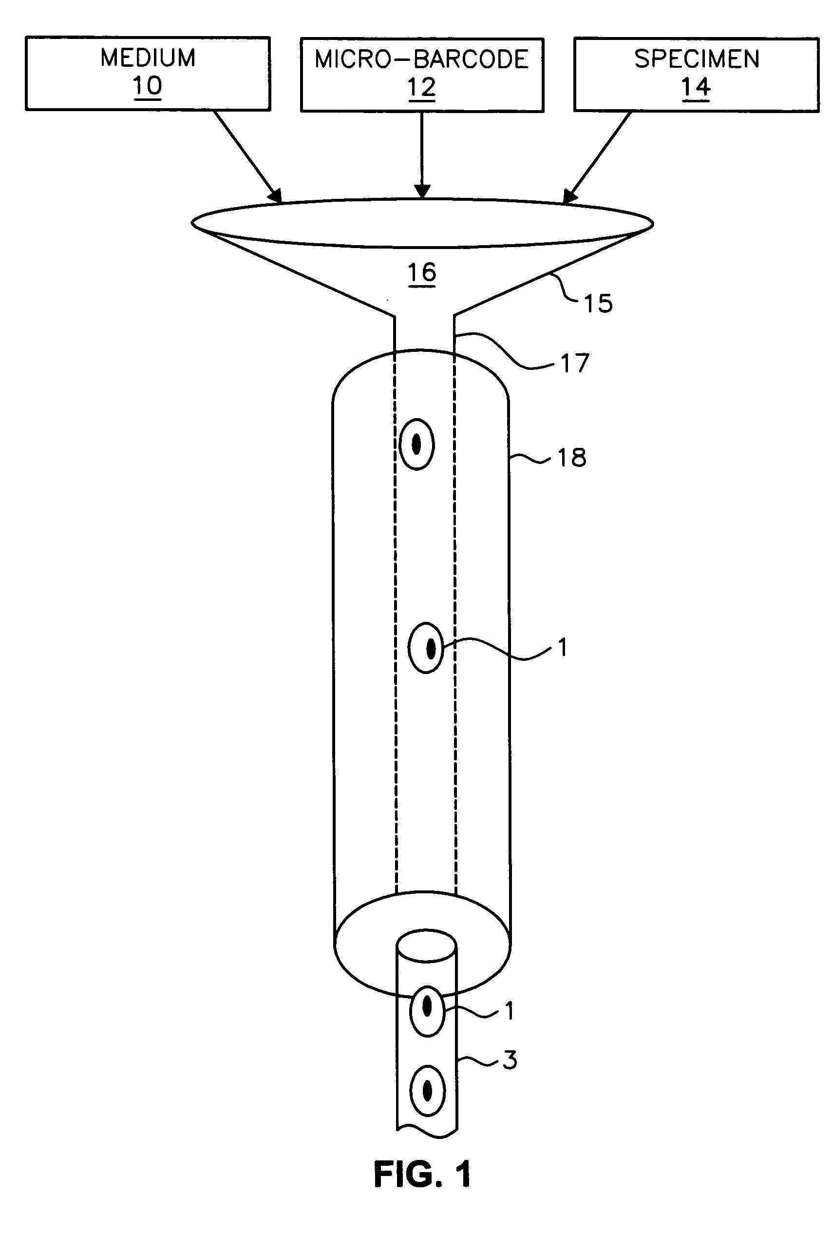
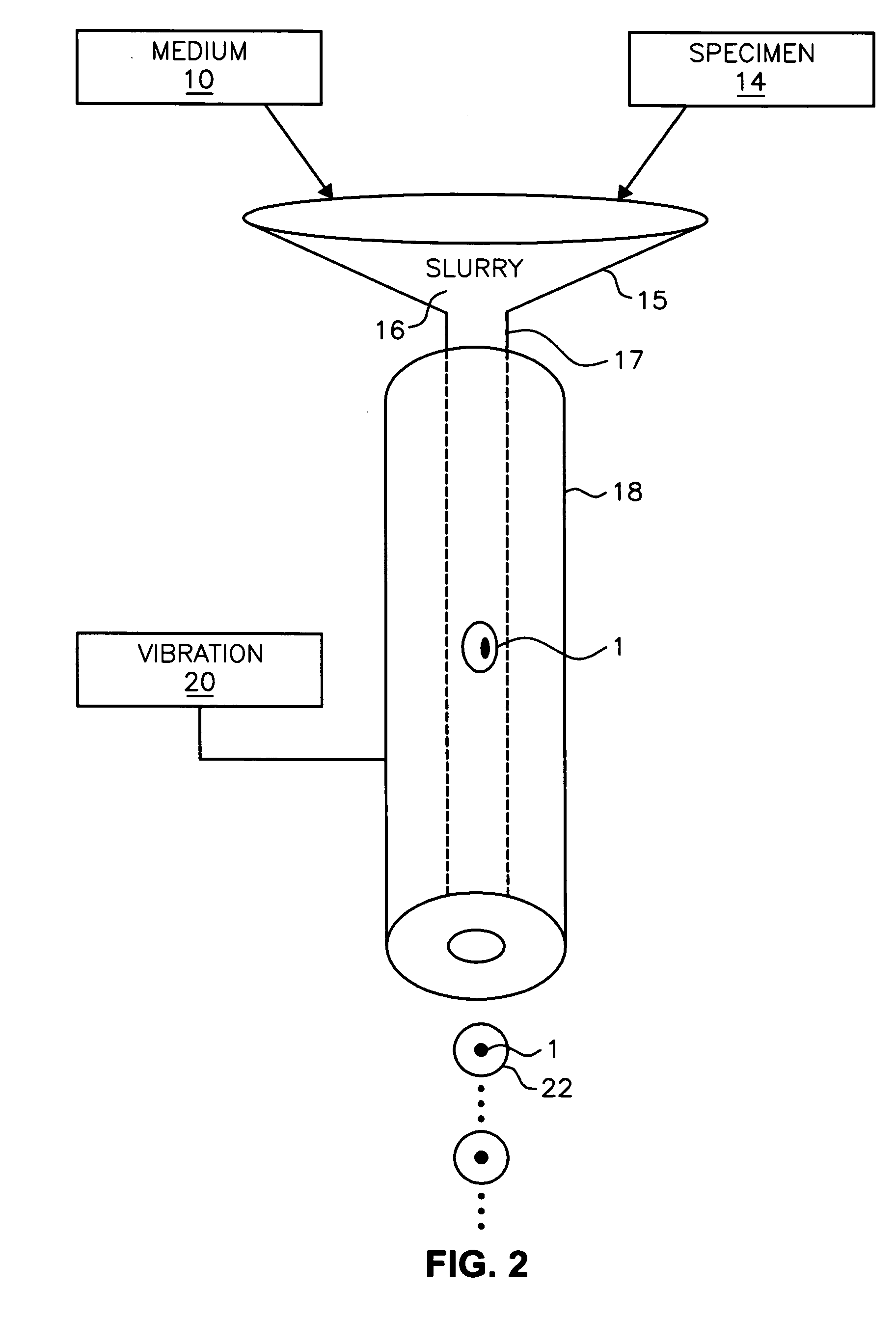
The present invention provides a method for embedding particles in a solid structure including the steps of extruding a slurry of particles and a polymeric solution into a linear polymer medium having particles embedded into a polymer portion; and curing the polymer portion of the linear polymer medium.

Description

RELATED APPLICATIONS [0001] This application claims the benefit of the priority date and is a continuation-in-part of co-pending U.S. patent application Ser. No. 10 / 126,026, filed Apr. 19, 2002, of Nelson entitled “VARIABLE-MOTION OPTICAL TOMOGRAPHY OF SPECIMEN PARTICLES,” the disclosure of which is incorporated herein by this reference. [0002] This application is also related to concurrently filed application to Fauver et al. entitled, “IMPROVEMENTS IN OPTICAL PROJECTION TOMOGRAPHY MICROSCOPE,” attorney docket no. 60097US that is assigned to the same assignees as the present application and the disclosure of which is hereby incorporated by reference.FIELD OF THE INVENTION [0003] The present invention relates generally to the field of specimen preparations and, more particularly, to a method for preparing cells in transport meddium such as a thixotropic gel or a polymer medium for use in three dimensional image acquisition. BACKGROUND OF THE INVENTION [0004] For some imaging applica...

Claims


Application Information

Patent Timeline
IPC IPC(8): A61B10/00G01N21/17G01N15/14G01N21/21G01N21/47
CPCG01N15/1468G01N15/147G01N15/1475G01N21/4795G06T2211/421G01N2015/1075G01N2201/0813G01N2223/612G06T11/006G01N2015/1006
Inventor FAUVER, MARK E.NELSON, ALAN C.RAHN, JOHN RICHARDSEIBEL, ERIC J.PATTEN, FLORENCE W.MCGUIRE, SHAWN
Owner UNIV OF WASHINGTON
Why Eureka
Try Eureka