[0008] The invention provides devices and methods for preventing
ischemic stroke in patients undergoing
percutaneous invasive vertebral or carotid procedures, including
angioplasty,
stent placement,
atherectomy, and / or filter
insertion, by reversing
blood flow down a vertebral artery, an extracranial or intracranial internal carotid artery, an
external carotid artery, and / or a
common carotid artery and into the ipsilateral subclavian artery. In this way, embolic debris generated as a result of placing
instrumentation within a diseased artery is diverted to the subclavian artery, thereby preventing stroke by minimizing distal embolization to the narrow cerebral vessels. The devices and methods are also useful to remove an
embolus and improve flow (by reversing collateral
blood flow across the circle of Willis) in patients with
acute stroke.
[0013] It will be understood that coarctation in the
aorta increases the pressure gradient from the left
cerebral arteries to the right
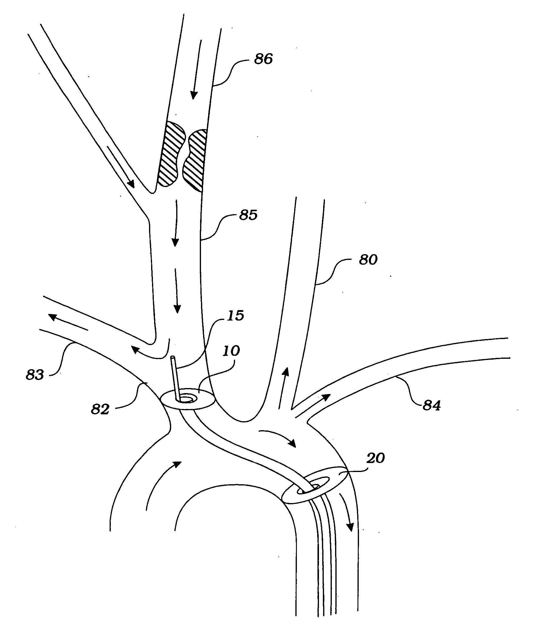
cerebral arteries, thereby enhancing flow reversal in the right cerebral arteries (including the right CCA, the right ICA, the right ECA, and the right vertebral artery). At a critically low brachiocephalic pressure downstream or distal to the
constriction, blood flow in the carotid and vertebral arteries is reversed to pass over the
atheromatous lesion and into the right subclavian artery. The flow reversal can be verified fluoroscopically with dye.
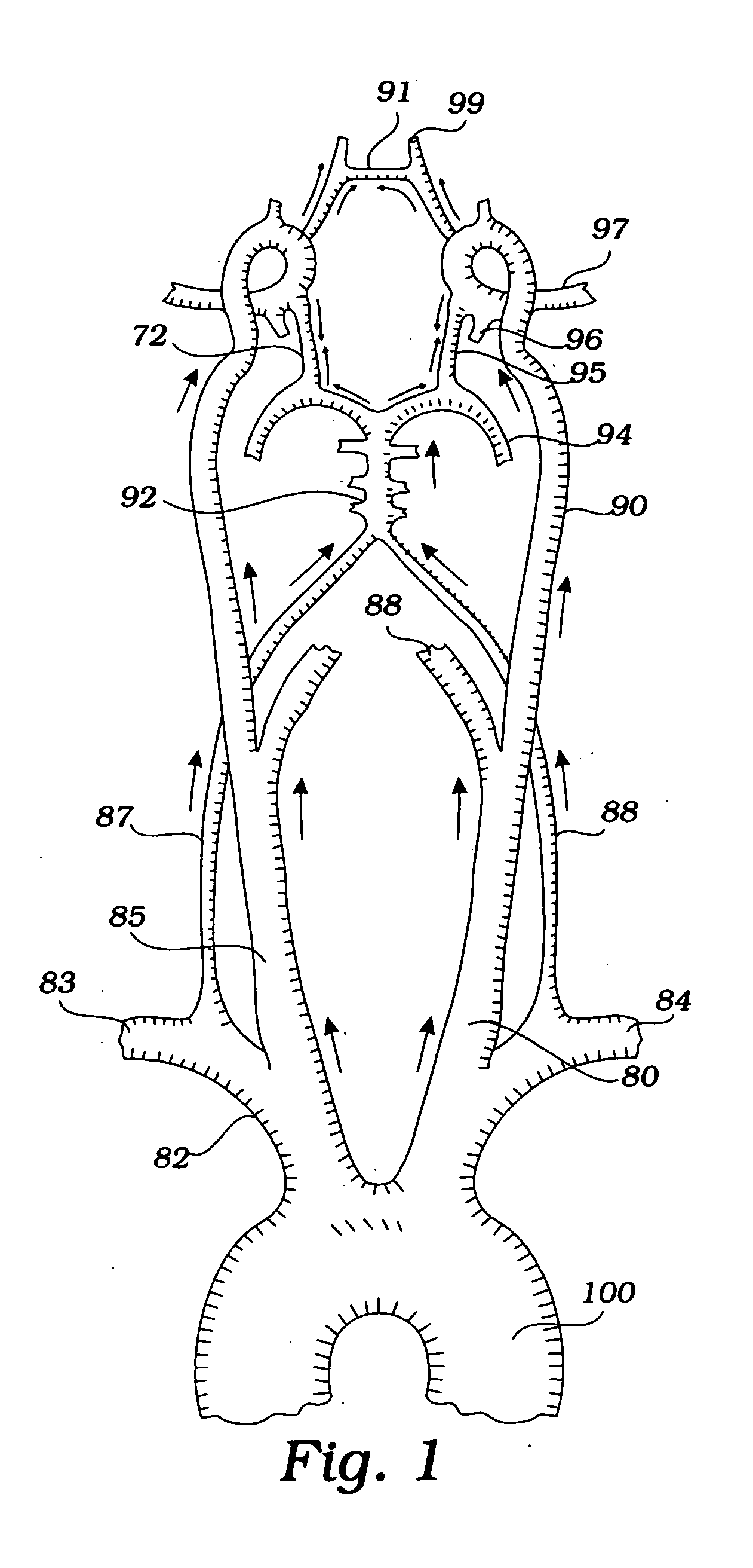
[0016] In another method, a coarctation constrictor is positioned in the
aorta upstream or downstream of the
left subclavian artery, and a second constrictor is positioned in the
left subclavian artery upstream of the
left vertebral artery. The second constrictor is expanded to reduce pressure downstream or distally in the
left subclavian artery. The coarctation constrictor is expanded to augment
cerebral blood flow to the right subclavian artery, the left CCA, the right brachiocephalic artery, and the right CCA. Coarctation in the aorta increases the pressure gradient from the right cerebral arteries to the
left vertebral artery, thereby enhancing flow reversal in the
left vertebral artery. At a critically low left subclavian pressure downstream or distal to the
constriction, blood flow in the left vertebral artery is reversed to pass over the
atheromatous lesion and into the left subclavian artery. The flow reversal can be verified fluoroscopically with dye.
[0017] In another method, a coarctation constrictor is positioned in the aorta upstream or downstream of the left subclavian artery, and a second constrictor is positioned in the left common carotid artery. The second constrictor is expanded to reduce pressure downstream or distally in the left common carotid artery. The coarctation constrictor is expanded to augment
cerebral blood flow to the left subclavian artery, the right brachiocephalic artery, and the right CCA. It will be understood that coarctation in the aorta increases the pressure gradient from the right cerebral arteries and left vertebral artery to the left CCA, thereby enhancing flow reversal in the left CCA.
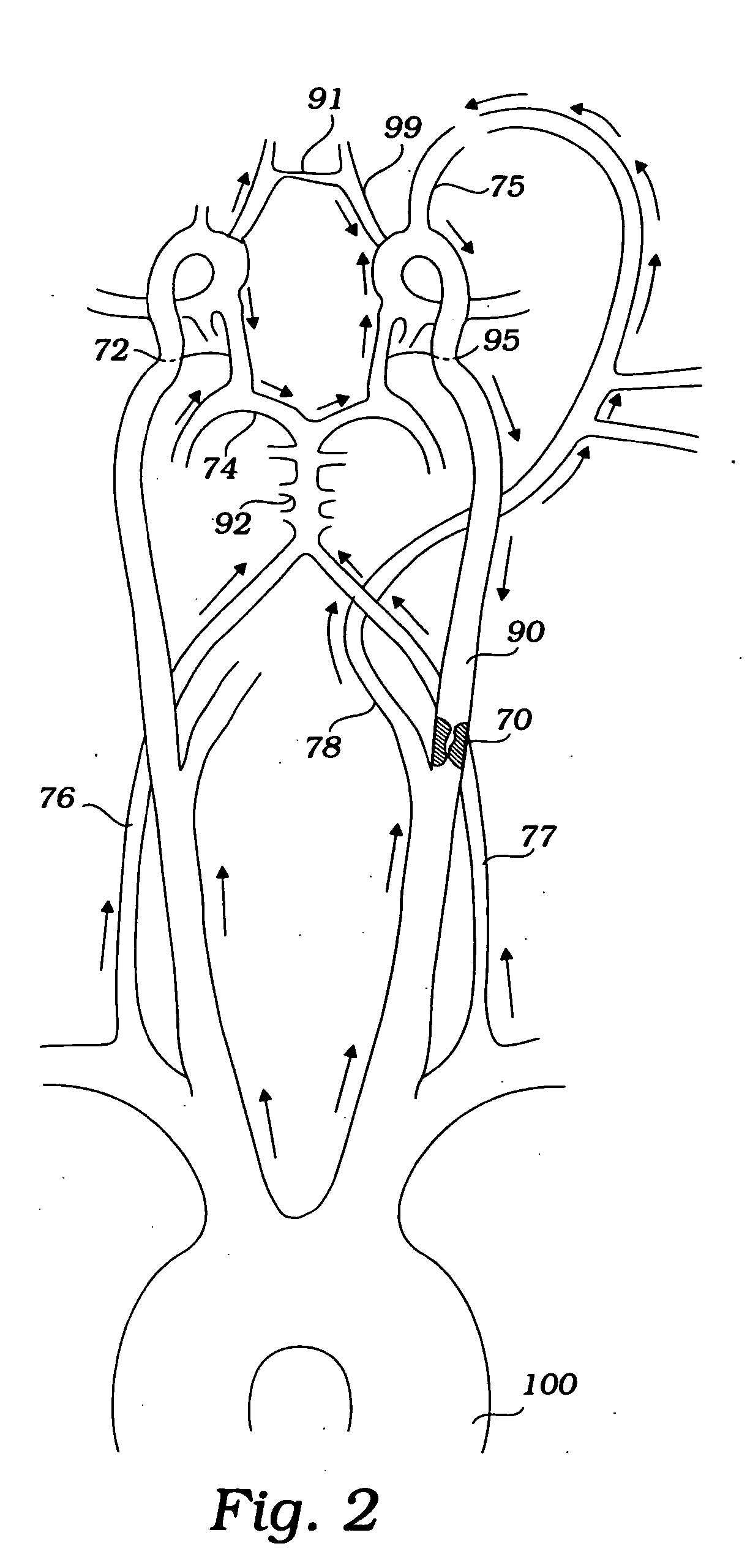
[0018] In another method, a coarctation constrictor is positioned in the aorta upstream or downstream of the left subclavian artery, and a second constrictor-occluder is positioned in the right common carotid artery or left common carotid artery.
Blood flow is reversed down the right internal carotid artery and into the right external carotid artery or down the left internal carotid artery and into the left external carotid artery, when the constrictors are expanded. A filter may be located in the external carotid artery to capture embolic debris. A third constrictor may be located in the external carotid artery to enhance the pressure gradient between the internal carotid artery and external carotid artery to enhance flow reversal in the internal carotid artery.
[0020] It will be understood that there are several advantages in using the devices and methods disclosed herein for prevention of distal embolization during use of
instrumentation in the
carotid arteries. For example, the devices (1) abolish the need for suction distal to the constricting / occluding member, thereby minimizing
blood loss, (2) eliminate the need for systemic anticoagulation, pumping, and a second arterial or venous stick, all of which are required where suction is employed, (3) can be used to introduce a variety of diagnostic or therapeutic instruments to the
carotid arteries, (4) can be used in any procedures that require instrumentation within the carotid artery, (5) can be used for definitive treatment of acute or subacute
ischemic stroke, (6) can be used in the angiogram or
fluoroscopy suite available in most hospitals, (7) usually require only one
incision site for entry, and (8) can be used to perform an interventional procedure without
distal protection (e.g., a distal filter), and without crossing the
lesion.

