Apparatus and method for phased subarray imaging

a subarray and imaging technology, applied in tomography, applications, instruments, etc., can solve the problems of inability to achieve similar 3d imaging systems using conventional hardware and methods, inability to scale well to achieve similar 3d imaging systems, and inability to achieve and the analog nature of front-end hardware has not experienced the same reduction in cost and size. , to achieve the effect of reducing the electronic complexity of the front-end ultrasound imaging system and low beam ra

Inactive Publication Date: 2005-05-12
THE BOARD OF TRUSTEES OF THE LELAND STANFORD JUNIOR UNIV
View PDF34 Cites 62 Cited by
  • Summary
  • Abstract
  • Description
  • Claims
  • Application Information

AI Technical Summary

Benefits of technology

[0010] In view of the above, it is a primary object of the present invention to provide an apparatus and method for phased subarray imaging, including image reconstruction, in an ultrasound imaging system. The phased subarray imaging of this invention provides a high-beam-rate image and allows a reduction in the front-end electronic complexity of the ultrasound imaging system.
[0011] These and numerous other objects and advantages of the present invention will become apparent upon reading the following description.
[0012] The objects and advantages of the present invention are secured by an apparatus and method for phased subarray (PSA) imaging. An array of transducers is divided into a set of subarrays each having of multiple adjacent elements. Energy is transmitted with a transmit focal length from a subarray and complex responses to this energy are received by the subarray. The active subarray is multiplexed across the full array of transducers. Each subarray is fired multiple times to acquire QS beams, each defined by a direction in beam space and a plurality of receive focal lengths, that constitute a low-resolution subarray image with a low beam rate. The low-beam-rate subarray images are interpolate

Problems solved by technology

Conventional hardware and methods used for 2D ultrasound systems do not scale well to achieve similar 3D imaging systems.
A first challenge one faces when implementing such a system is fabricating the transducer array with reasonable yields.
A second challenge caused by a large channel count for a 3D ultrasound system is implementing the highly parallel front-end electronics required.
Front-end hardware has become one of the most space- and power-consuming parts of a typical ultrasound imaging system.
Unfortunately, the analog nature of the front-end hardware has not experienced an equal reduction in cost and size.
High-end commercial ultrasound machines still house the analog and mixed-signal, front-end electronics within a base unit, requiring costly and bulky probe cables that contain dedicated coaxial transmission lines for each transducer element.
Modern 2D imaging systems require this complex set of front-end electronics because they typically use conventional full phased array (FPA) imaging, which requires that all array elements be simultaneously active during transmit and receive.
These electronics are the primary contributor to the bulk, cost, and power consumption of a typical ultrasound imaging system.
In addition to high front-end hardware complexity, the large number of received signals required to form each beam causes a significant increase in transmit beamformer 158 and receive beamformer 160 complexity.
The implementation of precision delay lines for beam steering also places a large burden on the beamforming hardware.
While electronic components continue to become smaller, faster, and cheaper, it is still not feasible to implement a full set of channels required for a 2D transducer array for

Method used


Image

  • Apparatus and method for phased subarray imaging
  • Apparatus and method for phased subarray imaging
  • Apparatus and method for phased subarray imaging

Examples


example 1

[0059] For the special case of a fixed number of adjacent elements in each subarray and a fixed overlap 534 in FIG. 5 equal to half the number of adjacent elements M1 528 or M2 530 along laternal directions 512 or 516 in each subarray, the filter for interpolation is merely a bandpass; no additional spectral modification in a restoration filter is required. For narrowband imaging, the bandpass is 1D. For wideband imaging, the bandpass is in general 2D. The final high-beam-rate PSA image is a linear combination of individual high-beam-rate subarray images. For a 2D cross-section in the plane defined by the azimuth angle θ1 518 or the elevation angle θ2 520, the weight b[k] applied to the high-beam-rate subarray image corresponding to kth subarray is given by b1⁡[k]=(K1+12)-k-(K1-12),

where k is between 0 and K1−1. For this geometry, and more generally for overlap 534 less than half the number of adjacent elements M1 528 or M2 530, the frame rate reduction will never exceed a factor ...

example 2

[0064]FIG. 8 illustrates the impact of restoration filters including the subarray weights on the high-beam-rate PSA coarray. FIG. 8 shows the coarray corresponding to a 1D lateral cross-section of 3D data in the plane defined by the azimuth angle θ1 518 or elevation angle θ2 520. In this example, N1 510 or N2 514=10, M1 528 or M2 530=6 and K1 or K2=3 in FIG. 5. Referring back to FIG. 8, coarrays 810, 812 and 814 each correspond to a subarray. Each coarray 810, 812 and 814 has 11 non-zero samples (2 M1 528−1 or 2M2 530−1). Without restoration filtering, the weighted sum of coarrays 810, 812 and 814 results in an unrestored PSA coarray (not shown) that is not suitable for producing a high-beam-rate image. One possible set of restoration filters 816, 818 and 820 that could be used to obtain a high-beam-rate PSA coarray 828 that is comparable to a FPA coarray are shown. In this illustration, all the weights are equal to 2, and are incorporated into the magnitudes of the restoration filt...

example 3

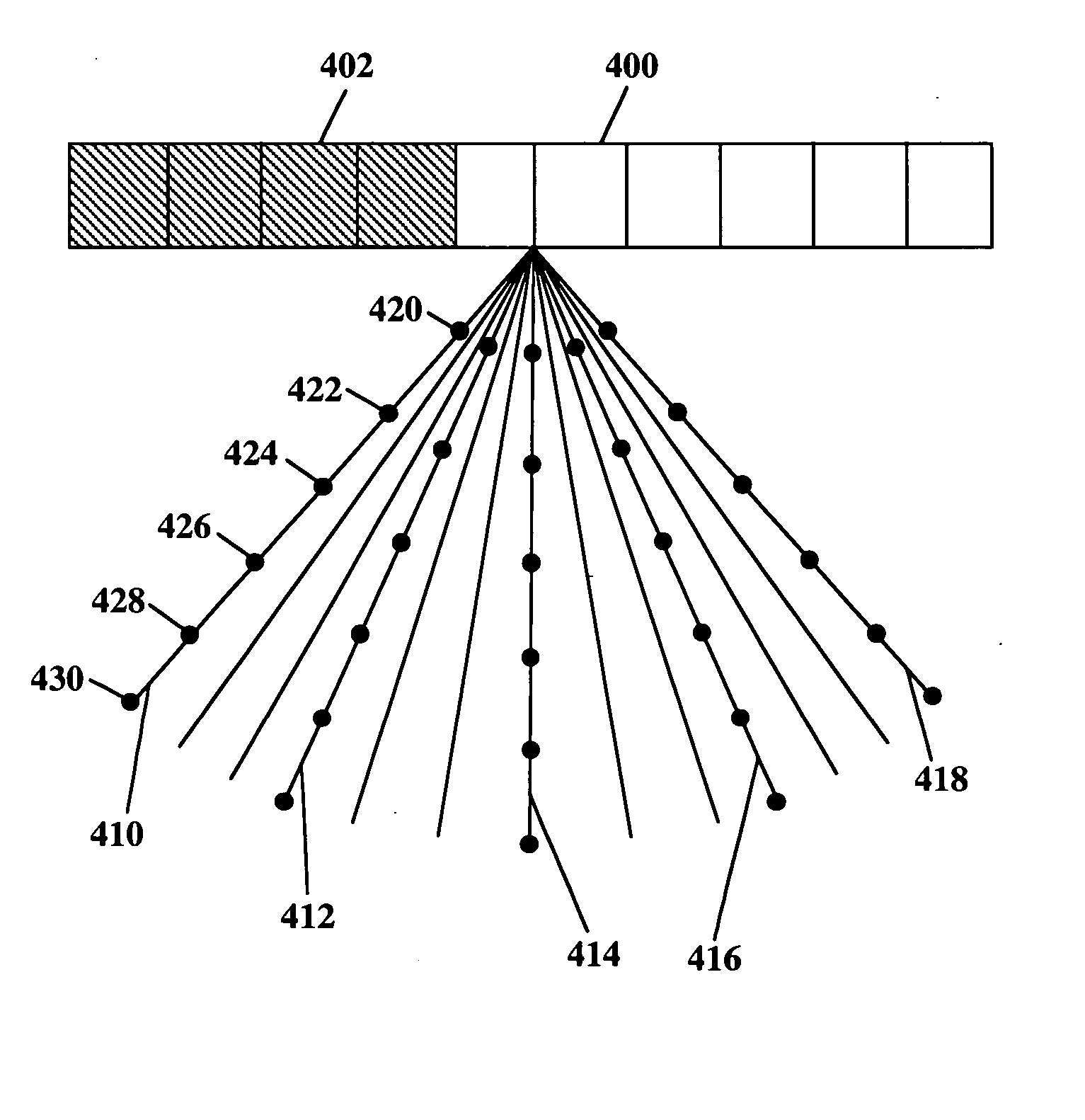
[0065] A comatrix serves as a useful tool for choosing which transmit and receive subarrays should be used to form the final coarray. Two example comatrices are shown in FIG. 9a-b, all with N1 510 or N2 514=16 and M1 528 or M2 530=4 (once again, FIG. 9a-b illustrates a 2D cross-section in the plane defined by the azimuth angle θ1 518 or elevation angle θ2 520 in FIG. 5). Each example demonstrates how different choices for the number of subarrays and the transmit / receive subarray combinations affect the restoration filter needed to achieve an FPA-comparable coarray.

[0066] For the example shown in FIG. 9a, the array 900 is divided into four non-overlapping, adjacent subarrays 910, 912, 914 and 916. Images are acquired using 16 permutations 918, 920, 922, 924, 926, 928, 930, 932, 934, 936, 938, 940, 942, 944, 946 and 948 of the subarrays 910, 912, 914 and 916, one for every transmit / receive combination. The weights used in summing the coarrays for permutations 918, 920, 922, 924, 926,...


PUM


Abstract

An invention for coherent array image formation and restoration is taught. The invention is applicable for both 2D and 3D imaging using either ID or 2D arrays, respectively. A transducer array is subdivided into subarrays, each subarray having a number of adjacent array elements. All elements of each subarray transmit and receive in parallel. The signals received from each subarray are delayed and summed to form scan lines, or beams. The low-beam-rate beams formed from each subarray are upsampled and interpolated prior to forming high-beam-rate images. Depending on the subarray geometry, a subarray-dependent restoration filter is also applied to the subarray beams. The restored beams from each subarray are combined to form the final high-beam-rate image. The invention significantly reduces the front-end hardware complexity compared to conventional methods such as full phased array imaging with comparable image quality.

Description

BACKGROUND OF THE INVENTION [0001] Real-time medical ultrasound imaging has played an increasingly important role in the diagnosis and treatment of disease. Ultrasound imaging is used for routine diagnostic procedures in obstetrics, gynecology, cardiology, and gastroenterology. The vast majority of ultrasound systems in use today provide two-dimensional (2D) cross-sections of the anatomy. While other imaging modalities such as magnetic resonance imaging and x-ray computed tomography have provided three-dimensional (3D) images since their inception, only recently have 3D ultrasound imaging systems become commercially available. These systems have the potential to revolutionize medical imaging by providing 3D visualization of the anatomy and blood flow in real-time. [0002] Conventional hardware and methods used for 2D ultrasound systems do not scale well to achieve similar 3D imaging systems. Modem 2D ultrasound scanners use a long 1D-transducer array having roughly 192 elements. Tran...

Claims


Application Information

Patent Timeline
IPC IPC(8): A61B8/14G01S7/52G01S15/89
CPCA61B8/14G01S7/52046G01S15/8915G01S15/8993G01S15/8927
Inventor JOHNSON, JEREMY A.KARAMAN, MUSTAFAKHURI-YAKUB, BUTRUS THOMAS
Owner THE BOARD OF TRUSTEES OF THE LELAND STANFORD JUNIOR UNIV
Why Eureka
Try Eureka